Category

Metal Impregnation, Non-Silver

Lynch & Inwood’s Gold for Amyloid

By Amyloid, Metal Impregnation, Metal Impregnation, Non-Silver, Protocols, Stain Target, Stain Type
Protocol

Lynch & Inwood's Gold

for Amyloid

7
steps
5
materials

Materials

  • Chloro-auric acid, 1% aqueous
  • Hydrogen peroxide, 3% aqueous
  • Iodine solution
    MaterialAmount
    Iodine1g
    Potassium iodide2g
    Distilled water100mL

    Mix the potassium iodide and iodine crystals together. Add 5 mL of the water and mix until both have dissolved. Add the rest of the water.

Tissue Sample

5-10µ sections from formalin fixed, paraffin embedded tissues, mounted on slides are suitable.


Protocol

  1. Bring sections to water through xylene and ethanols.
  2. Place in the iodine solution for 2½-5 minutes.
  3. Rinse with distilled water, 3 times of 5-10 seconds each.
  4. Place in chloro-auric acid solution for 2½-5 minutes.
  5. Rinse with distilled water, 3 times of 5-10 seconds each.
  6. Place in fresh 3% hydrogen peroxide at 37°C for 6-36 hours.
  7. Dehydrate with ethanol, clear with xylene and mount using a resinous medium.

Expected Results

  • Amyloid – golden yellow to faint purple.
  • Erythrocytes, muscle and liver – blue
  • Nuclei and cytoplasm – brown
  • Collagen – grey

Safety Note

Prior to handling any chemical, consult the Safety Data Sheet (SDS) for proper handling and safety precautions.


References

  1. Lynch, M.J. and Inwood, M.J.H., (1963),
    Gold as a permanent stain for amyloid,
    Stain Technology, v 38, page 260.

Schmorl’s Stain for Reducing Substances

By Intracytoplasmic Granules, Melanin & Enterochromaffin, Metal Impregnation, Metal Impregnation, Non-Silver, Protocols, Stain Target, Stain Type

Schmorl's Stain

for Reducing Substances

6
steps
4
materials

Materials

  • Nuclear fast red
  • Schmorl’s solution
    MaterialAmount
    Ferric chloride, 1% aqueous, fresh30mL
    Potassium ferricyanide, 1% aqueous, fresh4mL
    Distilled water6mL

This solution should be freshly prepared immediately before use.

Tissue Sample

5µ paraffin sections of neutral buffered formalin fixed tissue are suitable. Other fixatives are likely to be satisfactory.

Protocol

  1. Bring sections to distilled water via xylene and ethanol.
  2. Place into freshly made Schmorl’s solution for 10 minutes.
  3. Wash well with water.
  4. Counterstain with nuclear fast red.
  5. Rinse well with water.
  6. Dehydrate with ethanol, clear with xylene and mount with a resinous medium.

Expected Results

  • Reducing substances  –  blue
  • Nuclei  –  red

Notes

  • Reducing substances which may be present are colored blue. This includes melanin, for which it is a useful method, enterochromaffin and lipofuscin.
  • The Schmorl’s solution is from Lillie’s modification of Schmorl’s ferricyanide reduction method for tissue reducing substances.

Safety Note

Prior to handling any chemical, consult the Safety Data Sheet (SDS) for proper handling and safety precautions.

References

  1. Lillie, R.D., (1954)
    Histopathologic technique and practical histochemistry Ed.2
    Blakiston, New York, USA.

Perls’ Prussian Blue for Hemosiderin

By Metal Impregnation, Metal Impregnation, Non-Silver, Protocols, Stain Type

Perls' Prussian Blue

for Hemosiderin

7
steps
4
materials

Materials

  • Neutral red
  • Stock solution A
    MaterialAmount
    Potassium ferrocyanide2g
    Distilled water100mL
  • Stock solution B
    MaterialAmount
    Hydrochloric acid, conc.2mL
    Distilled water100mL
  • Working solution
    MaterialAmount
    Stock solution A1volume
    Stock solution B1volume

Make the working solution immediately before use. Do not use if more than 30 minutes old.

Tissue Sample

5 µ paraffin sections of neutral buffered formalin fixed tissue are suitable. Avoid iron containing materials and jars while fixing as these may contaminate the tissue. Acid containing fixatives may remove some of the iron deposits, but apart from that most are satisfactory.

Protocol

  1. Bring sections to distilled water with xylene and ethanol.
  2. Place into the working solution for 15 minutes.
  3. Rinse with distilled water, then tap water.
  4. Stain with neutral red for one minute.
  5. Rinse well with tap water.
  6. Dehydrate with ethanol.
  7. Clear with xylene.

Expected Results

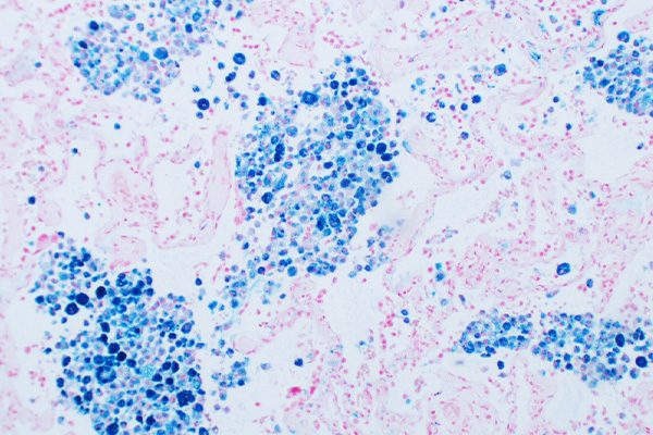
  • Ferric iron  –  blue
  • Nuclei  –  red

Notes

  • Avoid washing with tap water before placing into the working solution, as rust in the water or tap fixtures could cause false positive staining.
  • Wash well at step 3, as traces of iron will form a granular red deposit with neutral red.
  • Iron ores can be demonstrated, but the acid concentration in solution A may need to be increased to 10% or more.
  • Hemosiderin forms the body’s iron stores. The iron is in the ferric state, and may be demonstrated by releasing it from hemosiderin with hydrochloric acid, forming ferric chloride. The iron reacts with potassium ferrocyanide
    to form ferric ferrocyanide. This is an insoluble, blue compound known as Prussian blue or Berlin blue. The intensity of the colour gives some indication as to amount, but it is qualitative only. Other sources of ferric iron will also be demonstrated.
  • The reaction is: 4FeCl3 + 3K4Fe(CN)6 = Fe4[Fe(CN)6]3 + 12KCl

Safety Note

Prior to handling any chemical, consult the Safety Data Sheet (SDS) for proper handling and safety precautions.

References

  1. Culling, C.F.A., Allison, R.T. and Barr, W.T.
    Cellular Pathology Technique, Ed.4.
    Butterworth, London, UK.
  2. Susan Budavari, Editor, (1996)
    The Merck Index, Ed. 12
    Merck & Co., Inc., Whitehouse Station, NJ, USA

Howell’s Rubeanic Acid for Copper Deposits

By Metal Impregnation, Metal Impregnation, Non-Silver, Protocols, Stain Type

Howell's Rubeanic Acid

for Copper Deposits

7
steps
4
materials

Materials

  • Eosin Y, 0.1% in absolute ethanol.
  • Sodium acetate, 10% aqueous.
  • Stock rubeanic acid
    MaterialAmount
    Rubeanic acid50mg
    Ethanol, absolute50mL
  • Working rubeanic acid
    MaterialAmount
    Stock rubeanic acid2.5mL
    Sodium acetate, 10% aqueous.50mL

Tissue Sample

5 µ paraffin sections of neutral buffered formalin fixed tissue are suitable. Other fixatives are satisfactory.

Protocol

  1. Bring sections to water via xylene and ethanol.
  2. Place in the working rubeanic acid solution overnight at 37°C.
  3. Place into 70% ethanol for 15 minutes.
  4. Place into absolute ethanol for 6 hours.
  5. Lightly counterstain with alcoholic eosin.
  6. Rinse well with absolute ethanol.
  7. Clear with xylene and mount with a resinous medium.

Expected Results

  • Copper  –  green-black granules
  • Cytoplasm  –  pink

Notes

  • Use both negative and positive controls, as occasional failures in staining occur. A suitable positive control is a liver section from a known case of Wilson’s disease. Failing that, copper may be found in liver from chronic active hepatitis.
  • Copper forms a chelate with rubeanic acid, as do some other metals. Of these, cobalt and nickel may be confused with copper, so sodium acetate is added to the solution to inhibit them.
  • Rubeanic acid is also known as ethanedithioamide, dithiooxamide or dithiooxalic diamide, with the formulae below. It would appear that the copper is chelated between the sulphur and nitrogen (see Kiernan).

Rubeanic chemical structure

H2NCSCSNH2

Safety Note

Prior to handling any chemical, consult the Safety Data Sheet (SDS) for proper handling and safety precautions.

References

  1. Bancroft, J.D. and Stevens A. (1982)
    Theory and practice of histological techniques Ed. 2
    Churchill Livingstone, Edinburgh & London, UK.
  2. Susan Budavari, Editor, (1996)
    The Merck Index, Ed. 12
    Merck & Co., Inc., Whitehouse Station, NJ, USA
  3. Kiernan. J.A., (1999)
    Histological and histochemical methods: Theory and practice, Ed. 3
    Butterworth Heinemann, Oxford, UK.

Hale’s Colloidal Iron for Acid Mucosubstances

By Metal Impregnation, Metal Impregnation, Non-Silver, Protocols, Stain Type

Hale's Colloidal Iron

for Acid Mucosubstances

10
steps
9
materials

Using the colloidal iron suspension of Rhinehart and Abu’l Haj.

Materials

Solutions

  • Neutral red
  • Acetic acid, 2M (12%)
  • Potassium ferrocyanide, 2% aqueous
  • Hydrochloric acid, 2% aqueous
  • Rhinehart & Abu’l Haj’s colloidal iron suspension

Working colloidal iron

MaterialAmount
Colloidal iron suspension1vol
Acetic acid, 2M1vol

Perls’ solution

MaterialAmount
2% potassium ferrocyanide1vol
2% hydrochloric acid1vol

Tissue Sample

5µ paraffin sections of neutral buffered formalin fixed tissue are suitable. Other fixatives are usually satisfactory.

Protocol

  1. Bring sections to water via xylene and ethanol.
  2. Place into the working colloidal iron for 15-20 minutes.
  3. Wash with distilled water.
  4. Wash with running tap water for 5 minutes to remove all traces of colloidal iron
  5. Wash with distilled water.
  6. Place into freshly made Perls’ solution for 10 minutes.
  7. Wash with distilled water.
  8. Counterstain with neutral red for 1 minute.
  9. Dehydrate with ethanols.
  10. Clear with xylene and mount with a resinous medium.

Expected Results

  • Acid mucopolysaccharides  –  blue
  • Nuclei  –  red

Notes

  • The original method used a commercial colloidal iron preparation. This is still available. However, the colloidal iron suspension of Rhinehart and Abu’l Haj is reputed to produce a cleaner background. Other colloidal iron suspensions have also been recommended.
  • Since this method depends on the staining of iron compounds with the prussian blue reaction, any hemosiderin present will also be stained. If this is a concern, a control section should be stained which has not been treated with colloidal iron. Material stained blue in both sections should be discounted.
  • Nuclear fast red may also be used as a nuclear counterstain, or a Feulgen’s nucleal reaction may be applied before step 2, in which case the nuclear counterstain should be omitted.
  • A PAS may be applied following step 7, in which case the color of the nuclear counterstain should be changed, perhaps with a strictly progressive hemalum. Acid mucosubstances will be stained blue in contrast to red neutral mucosubstances. However, they are often present as mixtures and the contrast may not be clear.
  • Longley’s variant of this method includes a Feulgen’s nucleal reaction before step 2, and a Wiegert van Gieson counterstain following step 7, so that nuclei are black, cytoplasm is yellow and collagen red.

Safety Note

Prior to handling any chemical, consult the Safety Data Sheet (SDS) for proper handling and safety precautions.

References

  1. Culling C.F.A., (1963)
    Handbook of histopathological and histochemical techniques Ed. 2
    Butterworth, London, UK.
  2. Bancroft, J.D. and Stevens A. (1982)
    Theory and practice of histological techniques Ed. 2
    Churchill Livingstone, Edinburgh & London, UK.
  3. Lillie, R.D., (1954)
    Histopathologic technique and practical histochemistry Ed.2
    Blakiston, New York, USA.