Category

Stain Type

Lewis and Miller’s Trichrome for Pituitary cells

By Protocols, Stain Type, Trichrome Staining, Trichrome, Multi-Step

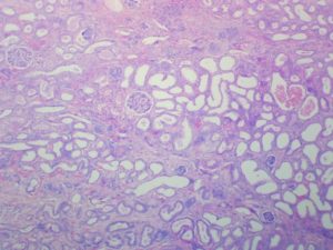
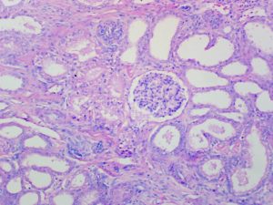
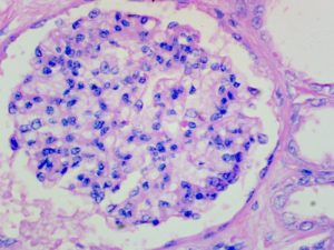
Lewis and Miller's Trichrome

for Pituitary cells

7
steps
5
materials

This is a modification of Kricheski’s method.

Materials

Solution A

MaterialAmount
Acid fuchsin0.25g
Distilled water100mL

Solution B

MaterialAmount
Methyl blue, 1% aqueous30mL
Orange G, 1% aqueous30mL
Phosphomolybdic acid, 1% aqueous30mL

Tissue Sample

5µ paraffin sections of neutral buffered formalin fixed tissue are likely suitable. Other fixatives are also likely to be satisfactory. Most trichrome stains benefit from picric acid or mercuric chloride fixation. Formalin fixed tissues may benefit from secondary fixation of sections in Bouin’s fluid.

Protocol

  1. Bring sections to water via xylene and ethanol.
  2. Place into solution A for 30 minute.
  3. Rinse well with distilled water.
  4. Place into solution B for 24 hours.
  5. Dip 2 or 3 times in 70% ethanol.
  6. Dehydrate and differentiate with ethanol for 1-3 minutes.
  7. Clear with xylene and mount with a resinous medium.

Expected Results

  • Nuclei  –  red
  • Acidophils  –  orange red
  • Basophils  –  blue
  • Chromophobes  –  Pale grey

Notes

  • Although not specified, an acid resistant nuclear stain such as Weigert’s iron hematoxylin could be inserted prior to staining with solution A.

Safety Note

Prior to handling any chemical, consult the Safety Data Sheet (SDS) for proper handling and safety precautions.

References

  1. Gray, Peter. (1954)
    The Microtomist’s Formulary and Guide.
    Originally published by: The Blakiston Co.
    Republished by: Robert E. Krieger Publishing Co.
    Citing:
    Kricheski, (1931)
    Stain technology, v.6, pp.97
    And:
    Lewis and Miller, (1938)
    Stain technology, v.14, pp.111

Lillie’s Alum Hematoxylin

By Dye Type, Hematoxylin and Eosin Staining, Mordanted Hematoxylin, Protocols, Stain Type

Lillie's Alum Hematoxylin

8
steps
6
materials

Materials

MaterialAmountFunction
Hematoxylin5 gDye
Ammonium alum50 gMordant
Distilled water700 mLSolvent
Glycerol300 mLStabilizer
Sodium iodate0.5 gOxidant
Glacial acetic acid20 mLAcidifier

Compounding Procedure

  1. Dissolve the dye and Alum in the water.
  2. Add the iodate, glycerol and acid.
  3. Leave overnight to ripen before using.

Protocol

  1. Bring sections to water with xylene and ethanol.
  2. Place into the staining solution for 5 minutes.
  3. Rinse well with water.
  4. Differentiate with acid ethanol if necessary.
  5. Rinse with water and blue.
  6. Rinse well with water.
  7. Counterstain if desired.
  8. Dehydrate with ethanol, clear with xylene and mount with a resinous medium.

Expected Results

  • Nuclei  –  blue
  • Background  –  as counterstain or unstained

Notes

  • This is a strong, regressive solution.
  • The staining time should be determined by trial.
  • Acid ethanol is 0.5% – 1% hydrochloric acid in 70% ethanol.
  • Blueing is done with alkaline solutions such as hard tap water, Scott’s tap water substitute, 0.1% ammonia water, 1% aqueous sodium acetate, 0.5% aqueous lithium carbonate etc.

Safety Note

Prior to handling any chemical, consult the Safety Data Sheet (SDS) for proper handling and safety precautions.

References

  1. Lillie, R.D.,
    Histopathologic Technic and Practical Histochemistry
    McGraw-Hill, New York, USA

Lillie & Earle’s Iron Hematoxylin

By Dye Type, Hematoxylin and Eosin Staining, Mordanted Hematoxylin, Protocols, Stain Type

Lillie & Earle's Iron Hematoxylin

8
steps
6
materials

Materials

Solution A

MaterialAmountFunction
Hematoxylin1 gDye
95% ethanol50 mLSolvent
Glycerol50 mLSolvent

Solution B

MaterialAmountFunction
Ferric ammonium sulfate15 gMordant
Ferrous sulfate15 gMordant
Distilled water100 mLSolvent

Compounding Procedure

  1. Make each solution separately.
  2. For use, combine equal parts of solutions A and B.
  3. The working solution may be used immediately.

Protocol

  1. Bring sections to water with xylene and ethanol.
  2. Place into the staining solution for an appropriate time.
  3. Rinse with tap water.
  4. If overstained, dip briefly in 1% hydrochloric acid in 70% ethanol.
  5. Wash well in running tap water to blue.
  6. Rinse with distilled water.
  7. Counterstain if desired.
  8. Dehydrate with ethanol, clear with xylene and mount with a resinous medium.

Expected Results

  • Nuclei  –  black
  • Background  –  as counterstain or unstained

Notes

  • The stock solutions are stable for some time.
  • The life of the working solution is not given. It is likely not stable for long, although the inclusion of glycerol and ferrous sulfate may improve its stability.
  • The staining time should be determined by trial, but 10-30 minutes should suffice.

Safety Note

Prior to handling any chemical, consult the Safety Data Sheet (SDS) for proper handling and safety precautions.

References

  1. Gray, Peter. (1954)
    The Microtomist’s Formulary and Guide.
    Originally published by: The Blakiston Co.
    Republished by: Robert E. Krieger Publishing Co.
    Citing:
    Lillie and Earle, (1937).
    American Journal of Pathology, v. 15, p. 765.

Lillie’s Iron Hematoxylin

By Dye Type, Hematoxylin and Eosin Staining, Mordanted Hematoxylin, Protocols, Stain Type

Lillie's Iron Hematoxylin

6
steps
6
materials

Materials

MaterialVariantFunction
19401954
Hematoxylin1 g0.5 gDye
Ferric chloride1.2 g1.2 gMordant
Ferrous sulfate4.44 gMordant
100% ethanol100 mLSolvent
Distilled water100 mL292 mLSolvent
Hydrochloric acid1 mL8 mLSolvent

Compounding Procedures

1940

    1. Dissolve the ferric chloride in half of the water.
    2. Dissolve the hematoxylin in the other half.

Combine and add the hydrochloric acid.

1954

  1. Dissolve the hematoxylin in the ethanol.
  2. Dissolve the ferric chloride and ferrous sulfate in the water.
  3. Add the hydrochloric acid.
  4. Combine the solutions.

Protocol

  1. Bring sections to water with xylene and ethanol.
  2. Place into the staining solution for 5-30 minutes.
  3. Wash well in running tap water to blue.
  4. Rinse with distilled water.
  5. Counterstain if desired.
  6. Dehydrate with ethanol, clear with xylene and mount with a resinous medium.

Expected Results

  • Nuclei  –  black
  • Background  –  as counterstain or unstained

Notes

  • The 1940 formula is likely not stable for long. If staining is too dark, dip briefly in 1% hydrochloric acid in 70% ethanol.
  • The 1954 formula is a modification of Wiegert’s iron hematoxylin. Lillie states that it is stable for several weeks with occasional use. It is progressive, and does not require differentiation.

Safety Note

Prior to handling any chemical, consult the Safety Data Sheet (SDS) for proper handling and safety precautions.

References

  1. Lillie, R.D., (1954)
    Histopathologic technique and practical histochemistry Ed.2
    Blakiston, New York, USA.
  2. Gray, Peter. (1954)
    The Microtomist’s Formulary and Guide.
    Originally published by: The Blakiston Co.
    Republished by: Robert E. Krieger Publishing Co.
    Citing:
    Lillie, (1940)
    Archiv für pathologische Anatomie,
    v. 29, p. 705.

Lillie’s Impregnation for Reticulin

By Metal Impregnation, Metal Impregnation, Silver, Protocols, Reticulin, Stain Target, Stain Type

Lillie's Impregnation

for Reticulin

20
steps
8
materials

Materials

  • Silver nitrate, 10% aqu.
  • Strong ammonium hydroxide (s.g. 0.88).
  • Potassium permanganate, 0.5% aqu.
  • Oxalic acid, 5% aqu.
  • Uranium nitrate, 1% aqu.
  • Formalin, 10% aqu.
  • Yellow gold chloride, 0.2% aqu.
  • Sodium thiosulphate, 5% aqu.
  • Neutral red, 1% aqu.

Preparation of Lillie’s Ammoniacal Silver

  1. Place 20 mL strong ammonium hydroxide in a flask.
  2. Add 10% silver nitrate drop by drop until the precipitate is almost dissolved (about 25-30 mL). The solution should have a faint opalescence.
  3. Dilute to twice its volume with distilled water.

Tissue Sample

5 µ paraffin sections of neutral buffered formalin fixed tissue are suitable. Other fixatives are likely to be satisfactory. A section adhesive is recommended.

Protocol

  1. Bring sections to water via xylene and ethanol.
  2. Oxidise with 0.5% potassium permanganate for 2 minutes.
  3. Rinse well with tap water.
  4. Bleach in Oxalic acid for a 2 minutes.
  5. Rinse well with tap water.
  6. Sensitise with 1% uranium nitrate solution for 5-10 seconds.
  7. Rinse with distilled water.
  8. Treat with ammoniacal silver solution for 3 minute.
  9. Rinse briefly with 90% ethanol.
  10. Rinse with distilled water.
  11. Place in 10% formalin for 2 minutes.
  12. Rinse well with tap water.
  13. Rinse with distilled water.
  14. Tone with 0.2% gold chloride solution.
  15. Rinse with distilled water.
  16. Fix in 5% sodium thiosulphate for 2 minute.
  17. Wash well with running tap water.
  18. Counterstain with neutral red for 1 minute.
  19. Rinse with tap water.
  20. Dehydrate with ethanol, clear with xylene, and mount with a resinous medium.

Expected Results

  • Reticulin fibres  –  black
  • Nuclei  –  red
  • Background  –  grey

Notes

  • Ensure that the ammonium hydroxide is fresh and full strength. Keep well stoppered when not in use. After removing the amount required immediately restopper the bottle.
  • Improperly made ammoniacal silver solutions can affect the quality of the impregnation. There should be a faint, persistent opalescence, with only a faint smell of ammonia.
  • It is sometimes difficult to obtain uranium nitrate, particularly if it requires international transportation.
  • Toning is a variable step. Untoned sections give dark brown reticulin fibres on a paler brown background. Many microscopists prefer to tone for about 15 seconds to produce brown-black reticulin fibres on a pale grey-brown background. Others tone longer (a few minutes) to produce black reticulin fibres on a grey background. Longer toning produces purple tones. Tone according to the personal preference of the microscopist reviewing the slides.

Safety Note

Prior to handling any chemical, consult the Safety Data Sheet (SDS) for proper handling and safety precautions.

References

  1. Gray, Peter. (1954)
    The Microtomist’s Formulary and Guide.
    Originally published by: The Blakiston Co.
    Republished by: Robert E. Krieger Publishing Co.

Llewellyn’s Mordant Blue 3 for Nuclei

By Dye Type, Hematoxylin Alternatives, Protocols, Stain Type

Llewellyn's Mordant Blue 3

for Nuclei

9
steps
8
materials

This is a substitute for alum hematoxylin in the H&E stain.

Materials

  • An aqueous or alcoholic eosin Y solution
  • Solution A
    MaterialVariant
    19741978a1978b
    Mordant blue 30.25g1g1g
    Iron alum, 10% aqu.40mL90mL5mL
    Sulphuric acid5mL
    Hydrochloric acid10mL
    Acetic acid50mL
    Distilled water955mL860mL985mL

    For all variants:

    1. Dilute the iron alum with the water.
    2. Add the mordant blue 3 and dissolve.
    3. Add the specified acid, mix and filter.

    The solutions are usable immediately and they have a shelf life of more than one year.

  • Solution B
    MaterialVariant
    Var AVar B
    Hydrochloric acid, conc1mL1mL
    Ethanol, 70%99mL
    Distilled water99mL
  • Solution C
    MaterialAmount
    Sodium acetate0.5g
    Distilled water100mL

Tissue Sample

5 µ paraffin sections of neutral buffered formalin fixed tissue are suitable. Other fixatives are likely to be satisfactory.

Protocol

  1. Bring sections to water via xylene and ethanol.
  2. Place into solution A for 5 minutes.
  3. Rinse well with tap water.
  4. Differentiate with a variant of solution B:
    1. For the 1974 formula: var A for 1-3 seconds or var B for 30 seconds.
    2. For the 1978a formula: var A for about ten seconds.
    3. For the 1978b formula: do not differentiate. Proceed to step 6.
  5. Rinse well with tap water.
  6. Blue with solution C for about 30 seconds.
  7. Rinse well with tap water.
  8. Counterstain with an eosin solution.
  9. Dehydrate with ethanol, clear with xylene and mount with a resinous medium.

Expected Results

  • Nuclei  –  blue
  • Cytoplasm  –  shades of pink

Human kidney stained progressively with mordant blue 3 and eosin Y at 40x magnification

Human kidney stained progressively with mordant blue 3 and eosin Y at 40x magnification

Human kidney stained progressively with mordant blue 3 and eosin Y at 100x magnification

Human kidney stained progressively with mordant blue 3 and eosin Y at 100x magnification

Human kidney stained progressively with mordant blue 3 and eosin Y at 400x magnification

Human kidney stained progressively with mordant blue 3 and eosin Y at 400x magnification

Notes

  • At times hematoxylin powder has become difficult, if not impossible, to buy. Due to the importance of this dye in making alum hematoxylin for H&E stains, several other dyes were investigated as substitutes. Mordant blue 3 was one of these, and Llewellyn published three variants of this substitute, both progressive and regressive.
  • The 1974 and 1978a formulae are regressive. The 1978b formula is progressive.
  • The sections will be blue after solution A. The blueing referred to in step 6 will deepen this, but it’s main purpose is to remove any pink staining due to unmordanted dye.
  • Iron alum is ferric ammonium sulphate:  FeNH4(SO4)2·12H2O. Use clean crystals.

Safety Note

Prior to handling any chemical, consult the Safety Data Sheet (SDS) for proper handling and safety precautions.

References

  1. Llewellyn, B. D., 1974
    Mordant blue3: A readily available substitute for hematoxylin
    in the routine hematoxylin and eosin stain.
    Stain Technology, v. 49, pp. 347.
  2. Llewellyn, B. D., 1978
    Improved nuclear staining with mordant blue 3 as a hematoxylin substitute
    Stain Technology, v. 53, pp. 73.

Lugol’s Alum Hematoxylin

By Dye Type, Hematoxylin and Eosin Staining, Mordanted Hematoxylin, Protocols, Stain Type

Lugol’s Alum Hematoxylin

6
steps
6
materials

Materials

Lugol’s Iodide

MaterialAmountFunction
Iodine2 gOxidant
Potassium iodide4 g
Distilled water100 mLSolvent

Hematoxylin

MaterialAmountFunction
95% ethanol100 mLSolvent
Hematoxylin10 gDye

Alum

MaterialAmountFunction
Potassium alum6 gMordant
Distilled water500 mLSolvent

Working solution

MaterialAmount
Hematoxylin1.5 mL
Lugol’s iodine1.5 mL
Alum100 mL

Compounding Procedure

  1. Make each solution as listed.
  2. Combine as directed above.
  3. Leave for 15 minutes before use.

Protocol

  1. Bring sections to distilled water with xylene and ethanol.
  2. Place into the staining solution for 5-10 minutes.
  3. Rinse with water and blue.
  4. Rinse well with water.
  5. Counterstain if desired.
  6. Dehydrate with ethanol, clear with xylene and mount with a resinous medium.

Expected Results

  • Nuclei  –  blue
  • Background  –  as counterstain or unstained

Notes

  • Read these comments on Lugol’s iodine.
  • The solution is usable for a minimum of one week.
  • The working solution will stain at least 100 slides.
  • Since the solution is not acidified, a distilled water rinse before staining will reduce alkaline carry over.
  • The authors named this solution Lugol’s Hematoxylin.
  • Blueing is done with alkaline solutions such as hard tap water, Scott’s tap water substitute, 0.1% ammonia water, 1% aqueous sodium acetate, 0.5% aqueous lithium carbonate etc.

Safety Note

Prior to handling any chemical, consult the Safety Data Sheet (SDS) for proper handling and safety precautions.

References

  1. Clarke, G. and Dodds, H. M., (1983)
    Lugol’s Hematoxylin.
    Stain Technology, v 58, Nº 4, p. 232

McFarlane’s Trichrome 1st Variant for Muscle and Collagen

By Protocols, Stain Type, Trichrome Staining, Trichrome, One-Step

McFarlane's Trichrome 1st Variant

for Muscle and Collagen

10
steps
9
materials

Materials

  • Weigert’s iron hematoxylin or equivalent
  • Solution A
    MaterialAmount
    Picric acid0.2g
    Acid fuchsin1g
    Aniline blue2g
    Phosphotungstic acid1g
    Acetic acid, glacial2mL
    Distilled water98mL
  • Solution B
    MaterialAmount
    Acetic acid, glacial2mL
    Distilled water100mL
  • Solution C
    MaterialAmount
    Picric acid0.25g
    Phosphotungstic acid2.5g
    Ethanol, 95%10mL
    Distilled water90mL

Tissue Sample

5µ paraffin sections of neutral buffered formalin fixed tissue are suitable. Other fixatives are likely to be satisfactory. Most trichrome stains benefit from picric acid or mercuric chloride fixation. Formalin fixed tissues may benefit from secondary fixation of sections in Bouin’s fluid.

Protocol

  1. Bring sections to water via xylene and ethanol.
  2. Stain nuclei with Weigert’s iron hematoxylin or equivalent.
  3. Place into solution A for 5 minute.
  4. Rinse with solution B.
  5. Rinse with distilled water.
  6. Place into solution C until differentiated.
  7. Rinse with distilled water.
  8. Wash with solution B.
  9. Dehydrate with ethanol.
  10. Clear with xylene and mount with a resinous medium.

Expected Results

  • Nuclei  –  black
  • Erythrocytes  –  yellow
  • Muscle  –  red
  • Cytoplasm  –  red
  • Collagen  –  blue

Notes

  • McFarlane also published two other, multi-step, trichrome methods. One is simpler than the other.
  • Solutions A and C require dry weights of picric acid. This could be provided from a saturated aqueous solution (1 g in 78 mL). Alternate formulas are:

    Solution A

    MaterialAmount
    Picric acid, sat. aqueous16mL
    Distilled water82mL
    Acetic acid, glacial2mL
    Acid fuchsin1g
    Aniline blue2g
    Phosphotungstic acid1g

    Solution C

    MaterialAmount
    Picric acid, sat. aqueous20mL
    Distilled water70mL
    Ethanol, 95%10mL
    Phosphotungstic acid2.5g

Safety Note

Prior to handling any chemical, consult the Safety Data Sheet (SDS) for proper handling and safety precautions.

References

  1. Gray, Peter. (1954)
    The Microtomist’s Formulary and Guide.
    Originally published by: The Blakiston Co.
    Republished by: Robert E. Krieger Publishing Co.
    Citing:
    McFarlane, (1944)
    Stain Technology, v. 19, pp. 29

McFarlane’s Trichrome 2nd Variant for Muscle and Collagen

By Protocols, Stain Type, Trichrome Staining, Trichrome, Multi-Step

McFarlane's Trichrome 2nd Variant

for Muscle and Collagen

12
steps
9
materials

Materials

  • Weigert’s iron hematoxylin or equivalent
  • Solution A
    MaterialAmount
    Picric acid0.2g
    Acid fuchsin0.8g
    Acetic acid, glacial2mL
    Distilled water98mL
  • Solution B
    MaterialAmount
    Acetic acid, glacial2mL
    Distilled water98mL
  • Solution C
    MaterialAmount
    Picric acid1g
    Phosphotungstic acid10g
    Ethanol, 95%40mL
    Distilled water60mL
  • Solution D
    MaterialAmount
    Aniline blue2.5mL
    Acetic acid, glacial2.5g
    Distilled water97.5mL
  • Solution E
    MaterialAmount
    Picric acid0.25g
    Phosphotungstic acid2.5g
    Ethanol, 95%10mL
    Distilled water90mL

Tissue Sample

5µ paraffin sections of neutral buffered formalin fixed tissue are suitable. Other fixatives are likely to be satisfactory. Most trichrome stains benefit from picric acid or mercuric chloride fixation. Formalin fixed tissues may benefit from secondary fixation of sections in Bouin’s fluid.

Protocol

  1. Bring sections to water via xylene and ethanol.
  2. Stain nuclei with Weigert’s iron hematoxylin or equivalent.
  3. Place into solution A for 5 minute.
  4. Rinse with solution B.
  5. Place into solution C for 5 minutes.
  6. Rinse with distilled water.
  7. Place into solution D for 5-10 minutes.
  8. Rinse with solution B.
  9. Place into solution E for 5 minutes.
  10. Wash with solution B.
  11. Dehydrate with ethanol.
  12. Clear with xylene and mount with a resinous medium.

Expected Results

  • Nuclei  –  black
  • Erythrocytes  –  yellow
  • Cytoplasm  –  red
  • Muscle  –  red
  • Collagen  –  blue

Notes

  • McFarlane also has another multi-step method and a one step method.
  • Solutions A, C and E require dry weights of picric acid. These could be provided from saturated aqueous (1 g in 78 mL) or ethanolic (1 g in 12 mL) solutions. Alternate formulas are:

    Solution A

    MaterialAmount
    Picric acid, sat. alc.16mL
    Distilled water82mL
    Acetic acid, glacial2mL
    Acid fuchsin0.8g

    Solution C

    MaterialAmount
    Picric acid, sat. alc.12mL
    Distilled water60mL
    Ethanol, 95%28mL
    Phosphotungstic acid10g

    Solution E

    MaterialAmount
    Picric acid, sat. alc.20mL
    Distilled water70mL
    Ethanol, 95%10mL
    Phosphotungstic acid2.5g

Safety Note

Prior to handling any chemical, consult the Safety Data Sheet (SDS) for proper handling and safety precautions.

References

  1. Gray, Peter. (1954)
    The Microtomist’s Formulary and Guide.
    Originally published by: The Blakiston Co.
    Republished by: Robert E. Krieger Publishing Co.
    Citing:
    McFarlane, (1944)
    Stain Technology, v. 19, pp. 29

McFarlane’s Trichrome 3rd Variant for Muscle and Collagen

By Protocols, Stain Type, Trichrome Staining, Trichrome, Multi-Step

McFarlane's Trichrome 3rd Variant

for Muscle and Collagen

14
steps
9
materials

Materials

  • Regaud’s iron hematoxylin or equivalent
  • Solution A
    MaterialAmount
    Picric acid1g
    Orange G0.25g
    Ethanol, 95%80mL
    Distilled water20mL
  • Solution B
    MaterialAmount
    Acid fuchsin0.25g
    Ponceau 2R0.25g
    Acetic acid, glacial1mL
    Distilled water99mL
  • Solution C
    MaterialAmount
    Acetic acid, glacial2mL
    Distilled water98mL
  • Solution D
    MaterialAmount
    Picric acid1g
    Phosphotungstic acid10g
    Ethanol, 95%80mL
    Distilled water20mL
  • Solution E
    MaterialAmount
    Aniline blue2.5g
    Acetic acid, glacial2.5mL
    Distilled water97.5mL
  • Solution F
    MaterialAmount
    Picric acid0.5g
    Phosphotungstic acid5g
    Ethanol, 95%20mL
    Distilled water801mL

Tissue Sample

5µ paraffin sections of neutral buffered formalin fixed tissue are suitable. Other fixatives are likely to be satisfactory. Most trichrome stains benefit from picric acid or mercuric chloride fixation. Formalin fixed tissues may benefit from secondary fixation of sections in Bouin’s fluid.

Protocol

  1. Bring sections to water via xylene and ethanol.
  2. Stain nuclei with Régaud’s iron hematoxylin, but do not differentiate with iron alum.
  3. Place into solution A until nuclei are differentiated.
  4. Wash with water until erythrocytes alone remain yellow.
  5. Place into solution B for 5-10 minutes.
  6. Rinse with solution C.
  7. Place into solution D for 5 minutes until differentiated.
  8. Rinse with solution C.
  9. Place into solution E for 10 minutes.
  10. Rinse with solution C.
  11. Place into solution F until differentiated.
  12. Wash with solution C.
  13. Dehydrate with ethanol.
  14. Clear with xylene and mount with a resinous medium.

Expected Results

  • Nuclei  –  black
  • Erythrocytes  –  yellow
  • Cytoplasm  –  red
  • Muscle  –  red
  • Collagen  –  blue

Notes

  • McFarlane also has another multi-step method and a one step method.
  • Solutions A, D and F require dry weights of picric acid. These could be provided from a saturated ethanolic (1 g in 12 mL) solution. Alternate formulas are:

    Solution A

    MaterialAmount
    Picric acid, sat. alc.12mL
    Ethanol, 95%68mL
    Distilled water20mL
    Orange G0.25g

    Solution D

    MaterialAmount
    Picric acid, sat. alc.12mL
    Ethanol, 95%68mL
    Distilled water20mL
    Phosphotungstic acid10g

    Solution F

    MaterialAmount
    Picric acid, sat. alc.6mL
    Ethanol, 95%14mL
    Distilled water80mL
    Phosphotungstic acid5g

Safety Note

Prior to handling any chemical, consult the Safety Data Sheet (SDS) for proper handling and safety precautions.

References

  1. Gray, Peter. (1954)
    The Microtomist’s Formulary and Guide.
    Originally published by: The Blakiston Co.
    Republished by: Robert E. Krieger Publishing Co.
    Citing:
    McFarlane, (1944)
    Stain Technology, v. 19, pp. 29