Protocol
Nuclear Fast Red
for Nuclei
Materials
Solution A
| Material | Amount | |
|---|---|---|
| Nuclear fast red | 1 | g |
| Potassium aluminum sulphate | 50 | g |
| Distilled water | 500 | mL |
Preparation
- Dissolve the dye and alum into the water.
- Boil for 5 minutes. Cool and filter.
Solution B
| Material | Amount | |
|---|---|---|
| Tartrazine | 1 | g |
| Distilled water | 1 | L |
Tissue Sample
5 µ paraffin sections of neutral buffered formalin fixed tissue are suitable. Other fixatives are likely to be satisfactory.
Protocol
- Perform the main staining method.
- Place into solution A for 5 to 10 minutes
- Rinse with water.
- Optionally, place into solution B for 30 seconds.
- Rinse with distilled water.
- Return to the main staining method, or
- Dehydrate with ethanol, clear with xylene and mount with a resinous medium.
Expected Results
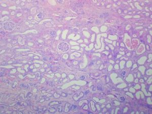
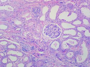
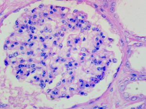
- Nuclei – red
- Cytoplasm – yellow or unstained
- Other tissues – according to the main staining method
Safety Note
Prior to handling any chemical, consult the Safety Data Sheet (SDS) for proper handling and safety precautions.
References
- Kiernan. J.A., (1999)
Histological and histochemical methods: Theory and practice, Ed. 3
Butterworth Heinemann, Oxford, UK.