Category

Protocols

[facetwp template="protocol_by_facet"]

Perls’ Prussian Blue for Hemosiderin

By Metal Impregnation, Metal Impregnation, Non-Silver, Protocols, Stain Type

Perls' Prussian Blue

for Hemosiderin
7
steps
4
materials

Materials

  • Neutral red
  • Stock solution A
    MaterialAmount
    Potassium ferrocyanide2g
    Distilled water100mL
  • Stock solution B
    MaterialAmount
    Hydrochloric acid, conc.2mL
    Distilled water100mL
  • Working solution
    MaterialAmount
    Stock solution A1volume
    Stock solution B1volume

Make the working solution immediately before use. Do not use if more than 30 minutes old.

Tissue Sample

5 µ paraffin sections of neutral buffered formalin fixed tissue are suitable. Avoid iron containing materials and jars while fixing as these may contaminate the tissue. Acid containing fixatives may remove some of the iron deposits, but apart from that most are satisfactory.

Protocol

  1. Bring sections to distilled water with xylene and ethanol.
  2. Place into the working solution for 15 minutes.
  3. Rinse with distilled water, then tap water.
  4. Stain with neutral red for one minute.
  5. Rinse well with tap water.
  6. Dehydrate with ethanol.
  7. Clear with xylene.

Expected Results

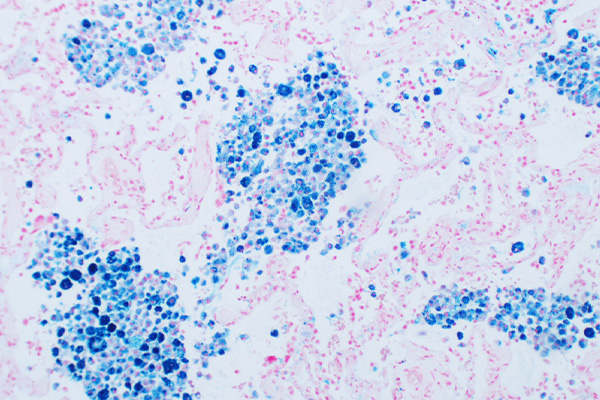
  • Ferric iron  –  blue
  • Nuclei  –  red

Notes

  • Avoid washing with tap water before placing into the working solution, as rust in the water or tap fixtures could cause false positive staining.
  • Wash well at step 3, as traces of iron will form a granular red deposit with neutral red.
  • Iron ores can be demonstrated, but the acid concentration in solution A may need to be increased to 10% or more.
  • Hemosiderin forms the body’s iron stores. The iron is in the ferric state, and may be demonstrated by releasing it from hemosiderin with hydrochloric acid, forming ferric chloride. The iron reacts with potassium ferrocyanide
    to form ferric ferrocyanide. This is an insoluble, blue compound known as Prussian blue or Berlin blue. The intensity of the colour gives some indication as to amount, but it is qualitative only. Other sources of ferric iron will also be demonstrated.
  • The reaction is: 4FeCl3 + 3K4Fe(CN)6 = Fe4[Fe(CN)6]3 + 12KCl

Safety Note

Prior to handling any chemical, consult the Safety Data Sheet (SDS) for proper handling and safety precautions.

References

  1. Culling, C.F.A., Allison, R.T. and Barr, W.T.
    Cellular Pathology Technique, Ed.4.
    Butterworth, London, UK.
  2. Susan Budavari, Editor, (1996)
    The Merck Index, Ed. 12
    Merck & Co., Inc., Whitehouse Station, NJ, USA

Lendrum, Slidders & Fraser’s Trichrome for Collagen and Other Tissues

By Protocols, Stain Type, Trichrome Staining, Trichrome, Multi-Step

Lendrum, Slidders & Fraser's Trichrome

for Collagen and Other Tissues

9
steps
6
materials

Materials

Small Molecular Weight (MW) Solution

MaterialAmount
Dye0.5g
Phosphotungstic acid2g
Ethanol, 95%100mL

Use one of the dyes from the appropriate list below:

Small MW Dyes

DyeCI NameAuthor’s Code
Martius yellowAcid yellow 24AY24
Orange GAcid orange 10A010
Azo-eosinAcid red 4AR4
Propalan red 3GXAcid red 57AR57
Acid fuchsinAcid violet 19AV19

Large MW solution

MaterialAmount
Dye0.5g
Acetic acid, glacial1mL
Distilled water99mL

Use one of the dyes from the appropriate list below:

Large MW Dyes

DyeCI NameAuthor’s Code
Sun yellowDirect yellow 11DY11
Sirius red F3BDirect red 80DR80
Benzo new blue GSDirect blue 10DR10
Durazol brilliant blue BDirect blue 109DB109

Tissue Sample

3-5µ paraffin sections of formal sublimate fixed tissue is preferred. If formalin fixation is used, the sections may require secondary fixation with picro-mercuric-alcohol.

Protocol

  1. Bring sections to water via xylene and ethanol.
  2. Stain nuclei with an acid resistant nuclear stain.
  3. Rinse with 95% ethanol.
  4. Place into small MW solution for 3 minutes.
  5. Rinse with distilled water.
  6. Place in the large MW solution for 30 minutes to 2 hours.
  7. Rinse with water.
  8. Dehydrate with absolute ethanol.
  9. Clear with xylene and mount with a resinous medium.

Expected Results

The results vary depending on the particular combination of small and large molecular weight dyes chosen. As a general guide, the small molecular weight dye can be expected to stain erythrocytes, cytoplasm and muscle. The large molecular weight dye can be expected to stain collagen. The authors particularly recommended the following combinations:

  • General use – AO10, DR80
  • After PAS – AY24, DY11
  • After silver – AR4, DY11

Notes

  • This technique has not gained favour as it uses some difficult to obtain dyes.
  • Picro-mercuric-alcohol is a saturated solution of both picric acid and mercuric chloride in absolute ethanol. It is usually applied to sections overnight after dewaxing and treating with ethanol, but before washing with water. If used, the iodine-thiosulphate sequence should be used to remove mercury pigment before staining.

Safety Note

Prior to handling any chemical, consult the Safety Data Sheet (SDS) for proper handling and safety precautions.

References

  1. Lendrum, A.C., Slidders, W. and Fraser, S., (1972),
    Renal hyalin: A study of amyloidosis and diabetic fibrinous vasculosis with new staining methods.,
    Journal of Clinical Pathology, v 25, page 373

Shoobridge’s Polychrome for Collagen and Other Structures

By Protocols, Stain Type, Trichrome Staining, Trichrome, Multi-Step

Shoobridge's Polychrome

for Collagen and Other Structures

21
steps
13
materials

Materials

  • Lillie’s alum hematoxylin
  • Iron alum
    MaterialAmount
    Ferric ammonium sulphate25g
    Distilled water500mL
    Glycerol70mL

    This should be pH 2.0.

  • Acid ethanol
    MaterialAmount
    Ethanol, absolute100mL
    Distilled water100mL
    Hydrochloric acid, concentrated1mL
  • Formal primer
    MaterialAmount
    Picric acid, saturated aqueous400mL
    Formalin, concentrated100mL

    This should be pH 2.0.

  • Naphthol yellow primer
    MaterialAmount
    Naphthol yellow S1g
    Distilled water400mL

    Adjust to pH 2.0 with HCl or NAOH.

  • Tungsto-orange
    MaterialAmount
    Phosphotungstic acid0.5g
    Distilled water200mL
    Orange G1g

    Preparation

    1. Dissolve the phosphotungstic acid.
    2. Then add the orange G.
    3. Adjust to pH 2.5 with HCl or NaOH.
  • Tungsto-acid fuchsin
    MaterialAmount
    Phosphotungstic acid0.5g
    Distilled water200mL
    Acid fuchsin1g

    Preparation

    1. Dissolve the phosphotungstic acid.
    2. Then add the acid fuchsin.
    3. Adjust to pH 2.5 with HCl or NaOH
  • Tungsto-methyl blue
    MaterialAmount
    Phosphotungstic acid0.5g
    Distilled water200mL
    Methyl blue1g

    Preparation

    1. Dissolve the phosphotungstic acid.
    2. Then add the acid fuchsin.
    3. Adjust to pH 2.5 with HCl or NaOH

Tissue Sample

5µ paraffin sections of tissues fixed in neutral buffered formalin for 6-24 hours as the primary fixative, followed by secondary fixation in 0.5% aqueous mercuric chloride for 30 minutes at 37°C, or for several hours at room temperature. Treatment with mercuric chloride may be omitted but the staining results will be inferior.

Protocol

  1. Bring sections to water via xylene and ethanol.
  2. Remove mercury pigment and wash well with tap water.
  3. Place into iron alum for 10 minutes.
  4. Rinse well with tap water.
  5. Rinse with distilled water.
  6. Place into Lillie’s hemalum for 10 minutes.
  7. Rinse well with tap water.
  8. Differentiate with acid ethanol for 15-30 seconds.
  9. Blue with running tap water (50°C). Avoid alkaline blueing solutions.
  10. Rinse with distilled water (50°C).
  11. Stain with naphthol yellow primer or picro-formal primer for 30 minutes at 50°C.
    Prewarm the solution.
  12. Wash in running tap water until differentiated (about 5 minutes at pH 6.5-7.0). Only erythrocytes, muscle and fibrin should be yellow.
  13. Stain in tungsto-orange solution for 5 minutes at room temperature.
  14. Rinse with tap water (pH 7.0 or less) for 15-30 seconds.
  15. Stain with tungsto-acid fuchsin for 5 minutes at room temperature.
  16. Rinse with tap water (pH 7.0 or less) for 15-30 seconds.
  17. Stain with tungsto-methyl blue for 5 minutes at room temperature.
  18. Rinse with tap water (pH 7.0 or less) for 15-30 seconds.
  19. Drain excess water.
  20. Dehydrate rapidly with 3 changes of absolute ethanol.
  21. Clear with xylene and mount with a resinous medium.

Expected Results

  • Erythrocytes  –  Yellow
  • Striated muscle  –  Yellow, or bright red if the primer is omitted
  • Fibrin  –  Varies: unstained, yellow-red, scarlet, magenta, blueish-red as it ages.
  • Fibrinoid  –  Varies: yellow-red, scarlet, crimson.
  • Albumin  –  Bright orange-red
  • Elastin  –  Clear pink-red
  • Nucleoli  –  Bright red
  • Platelets: Fresh to medium  –  Unstained to brick red
  • Platelets: Old  –  Purplish, grey or pale blue
  • Secretory granules (e.g. pituitary)  –  Yellow, red or blue
  • Thyroid colloid  –  Red or blue (inactive tends to be blue)
  • Bile  –  Emerald to olive green
  • Primary amyloid  –  Smoky blue
  • Secondary amyloid  –  Pale blue
  • Most mucins  –  Pale blue
  • Reticulin  –  Blue
  • Collagen  –  Deep Blue
  • Basement membrane  –  Intense deep Blue

Notes

  • It is strongly advised that the original paper be consulted as it contains a great deal of precise information.
  • The naphthol yellow primer was recommended for routine use, as the picro-formal primer gave redder results.
  • The original paper includes a one-step method using the same dyes. Three stock solutions are prepared. Each solution contains 2 g of dye dissolved into 100 mL of 1% phosphotungstic acid. The staining solution is made by combining equal parts of these three solutions. Staining times are not given and should be established by trial and error.

Safety Note

Prior to handling any chemical, consult the Safety Data Sheet (SDS) for proper handling and safety precautions.

References

  1. Shoobridge, Michael P. K., (1983),
    A new principle in polychrome staining: A system of automated staining, complementary to hematoxylin and eosin, and usable as a research tool.,
    Stain Technology, v 58, page 245

Cook’s Copper Hematoxylin

By Dye Type, Hematoxylin and Eosin Staining, Mordanted Hematoxylin, Protocols, Stain Type

Cook’s Copper Hematoxylin

7
steps
4
materials

Materials

MaterialAmountFunction
Hematoxylin10 gDye
Cupric sulfate2.5 gMordant
Potassium aluminum sulfate15 gMordant
Distilled water100 mLSolvent

Compounding procedure

  1. Place the dry powders in a mortar and grind together.
  2. Add sufficient water to make a paste.
  3. Leave for 48 hours, then add the remaining water.
  4. Leave for a further 12 hours, and filter.

Protocol

  1. Bring sections to water with xylene and ethanol.
  2. Place into the staining solution for an appropriate time.
  3. Rinse with water.
  4. Differentiate if necessary
  5. Wash well with water.
  6. Counterstain if wished.
  7. Dehydrate with ethanol, clear with xylene and mount with a resinous medium.

Expected Results

  • Nuclei  –  blue-black
  • Background  –  as counterstain or unstained

Notes

  • The staining time should be determined by trial.
  • The original method called for 15 grams of “extract of logwood”. Gray comments that this has about 70% dye content, so it has been specified to use 10 grams of dye.

Safety Note

Prior to handling any chemical, consult the Safety Data Sheet (SDS) for proper handling and safety precautions.

References

  1. Gray, Peter. (1954)
    The Microtomist’s Formulary and Guide.
    Originally published by: The Blakiston Co.
    Republished by: Robert E. Krieger Publishing Co.
    Citing:
    Hogg, J. (1883)
    The microscope
    Routledge, London, England

Howell’s Rubeanic Acid for Copper Deposits

By Metal Impregnation, Metal Impregnation, Non-Silver, Protocols, Stain Type

Howell's Rubeanic Acid

for Copper Deposits

7
steps
4
materials

Materials

  • Eosin Y, 0.1% in absolute ethanol.
  • Sodium acetate, 10% aqueous.
  • Stock rubeanic acid
    MaterialAmount
    Rubeanic acid50mg
    Ethanol, absolute50mL
  • Working rubeanic acid
    MaterialAmount
    Stock rubeanic acid2.5mL
    Sodium acetate, 10% aqueous.50mL

Tissue Sample

5 µ paraffin sections of neutral buffered formalin fixed tissue are suitable. Other fixatives are satisfactory.

Protocol

  1. Bring sections to water via xylene and ethanol.
  2. Place in the working rubeanic acid solution overnight at 37°C.
  3. Place into 70% ethanol for 15 minutes.
  4. Place into absolute ethanol for 6 hours.
  5. Lightly counterstain with alcoholic eosin.
  6. Rinse well with absolute ethanol.
  7. Clear with xylene and mount with a resinous medium.

Expected Results

  • Copper  –  green-black granules
  • Cytoplasm  –  pink

Notes

  • Use both negative and positive controls, as occasional failures in staining occur. A suitable positive control is a liver section from a known case of Wilson’s disease. Failing that, copper may be found in liver from chronic active hepatitis.
  • Copper forms a chelate with rubeanic acid, as do some other metals. Of these, cobalt and nickel may be confused with copper, so sodium acetate is added to the solution to inhibit them.
  • Rubeanic acid is also known as ethanedithioamide, dithiooxamide or dithiooxalic diamide, with the formulae below. It would appear that the copper is chelated between the sulphur and nitrogen (see Kiernan).

Rubeanic chemical structure

H2NCSCSNH2

Safety Note

Prior to handling any chemical, consult the Safety Data Sheet (SDS) for proper handling and safety precautions.

References

  1. Bancroft, J.D. and Stevens A. (1982)
    Theory and practice of histological techniques Ed. 2
    Churchill Livingstone, Edinburgh & London, UK.
  2. Susan Budavari, Editor, (1996)
    The Merck Index, Ed. 12
    Merck & Co., Inc., Whitehouse Station, NJ, USA
  3. Kiernan. J.A., (1999)
    Histological and histochemical methods: Theory and practice, Ed. 3
    Butterworth Heinemann, Oxford, UK.

Faure’s Iron Hematoxylin

By Dye Type, Hematoxylin and Eosin Staining, Mordanted Hematoxylin, Protocols, Stain Type

Faure's Iron Hematoxylin

8
steps
6
materials

Materials

Solution A

MaterialAmountFunction
Hematoxylin3.2 gDye
90% ethanol100 mLSolvent

Solution B

MaterialAmountFunction
Ferric chloride0.2 gMordant
Cupric acetate0.1 gMordant
Distilled water100 mLSolvent
Hydrochloric acid2 mLAcidifier

Compounding procedure

  1. Make each solution seperately.
  2. For use, combine equal parts of solutions A and B.
  3. The working solution may be used immediately, but is not stable for long.

Protocol

  1. Bring sections to water with xylene and ethanol.
  2. Place into the staining solution for 5 seconds.
  3. Rinse with tap water.
  4. Dip briefly in 1% hydrochloric acid in 70% ethanol.
  5. Wash well in running tap water to blue.
  6. Rinse with distilled water.
  7. Counterstain if desired.
  8. Dehydrate with ethanol, clear with xylene and mount with a resinous medium.

Expected Results

  • Nuclei  –  black
  • Background  –  as counterstain or unstained

Notes

  • The stock solutions are stable for some time.
  • The working solution should be made fresh.
  • This method is suitable as an acid resistant nuclear stain.
  • Only a very short staining time of 5 seconds is required.
  • The solution incorporates two mordants, iron and copper.

Safety Note

Prior to handling any chemical, consult the Safety Data Sheet (SDS) for proper handling and safety precautions.

References

  1. Gray, Peter. (1954)
    The Microtomist’s Formulary and Guide.
    Originally published by: The Blakiston Co.
    Republished by: Robert E. Krieger Publishing Co.
    Citing:
    Faure, (1929)
    Compte rendu hebdomadaire des séances et mémoires de la Société de biologie,
    v.90, p.87.

Cresyl Violet Stain for Nissl Bodies

By Intracytoplasmic Granules, Nissl Bodies, Protocols, Stain Target

Cresyl Violet Stain

for Nissl Bodies

11
steps
4
materials

Materials

Staining Solution

MaterialAmount
Cresyl violet1g
Distilled water100mL

Differentiator – Option 1

MaterialAmount
Ethanol, 95%100mL
Cajeput oila few drops

Differentiator – Option 2

MaterialAmount
Ethanol, 95%100mL
Acetic acid, glacial5 drops

Tissue Sample

5µ paraffin sections of neutral buffered formalin fixed tissue are suitable. Other fixatives, particularly if ethanolic, are likely to be satisfactory.

Protocol

  1. Bring sections to water via xylene and ethanol.
  2. Place into the staining solution for 15-30 minutes.
  3. Rinse with water, dehydrate with absolute ethanol and clear with xylene.
  4. Leave in xylene for one hour.
  5. Rinse well with absolute ethanol.
  6. Bring one slide at a time to 95% ethanol.
  7. Differentiate in either cajeput ethanol or acetic ethanol, controlling microscopically.
  8. Rinse well with 95% ethanol to stop differentiation.
  9. Dehydrate with absolute ethanol.
  10. Clear with xylene
  11. Coverslip using a resinous medium.

Expected Results

  • Nissl bodies  –  blue-violet
  • Nuclei  –  blue violet

Notes

  • The first dehydration and clearing of the stained section (steps 3-4) improves the sharpness of diffentiation and is recommended.
  • Differentiation, as in steps 5-10, may be repeated if necessary.

Safety Note

Prior to handling any chemical, consult the Safety Data Sheet (SDS) for proper handling and safety precautions.

References

  1. Drury, R A, and Wallington, E A, (1967).
    Carleton’s histological technique., Ed. 5., pp. 379.
    Oxford University Press, London, England.
  2. Bench Manual

Crossman’s Trichrome for Muscle and Collagen

By Protocols, Stain Type, Trichrome Staining, Trichrome, Multi-Step
Protocol

Crossman's Trichrome

for Muscle and Collagen

12
steps
7
materials

Materials

  • Solution A
    MaterialAmount
    Acid fuchsin0.3g
    Orange G2g
    Acetic acid, glacial1mL
    Distilled water100mL
    Thymola small crystal
  • Solution B
    MaterialAmount
    Phosphomolybdic acid1g
    Distilled water100mL
  • Solution C
    • Option 1
      MaterialAmount
      Aniline blue2g
      Acetic acid, glacial2mL
      Distilled water98mL
    • Option 2
      MaterialAmount
      Light green SF1g
      Acetic acid, glacial1mL
      Distilled water99mL
  • Solution D
    MaterialAmount
    Acetic acid, glacial1mL
    Distilled water99mL

Tissue Sample

5µ paraffin sections of neutral buffered formalin fixed tissue are suitable. Other fixatives are likely to be satisfactory. Most trichrome stains benefit from picric acid or mercuric chloride fixation.


Protocol

  1. Bring sections to water via xylene and ethanol.
  2. Stain nuclei with an acid resistant nuclear stain such as Weigert’s iron hematoxylin.
  3. Place into solution A for 1 minute.
  4. Rinse with distilled water.
  5. Place into solution B until collagen is decolorised.
  6. Rinse quickly with distilled water.
  7. Place into solution C for 5 minutes.
  8. Rinse with distilled water.
  9. Place into solution D until differentiated.
  10. Rinse with distilled water.
  11. Dehydrate with ethanol.
  12. Clear with xylene and mount with a resinous medium.

Expected Results

  • Nuclei  –  black
  • Erythrocytes  –  yellow
  • Cytoplasm  –  red
  • Collagen  –  blue or green

Safety Note

Prior to handling any chemical, consult the Safety Data Sheet (SDS) for proper handling and safety precautions.


References

  1. Gray, Peter. (1954)
    The Microtomist’s Formulary and Guide.
    Originally published by: The Blakiston Co.
    Republished by: Robert E. Krieger Publishing Co.

Csaba’s Stain for Mast Cells

By Intracytoplasmic Granules, Mast Cells, Protocols, Stain Target

Csaba's Stain

for Mast Cells

5
steps
6
materials

Materials

Staining solution

MaterialAmount
Alcian blue0.36g
Safranin0.18g
Ferric ammonium sulfate0.48g
Walpole’s acetate HCl buffer100mL

Walpole’s acetate-HCl buffer, pH 1.42

MaterialAmount
Sodium acetate, M/1100mL
Hydrochloric acid, M/1120mL

Tissue Sample

Paraffin sections of neutral buffered formalin fixed tissue are suitable. Mercuric chloride fixatives are reputed to emphasise metachromasia. Other fixatives may be satisfactory.

Protocol

  1. Bring sections to water via xylene and ethanol.
  2. Place in to staining solution for 10-20 minutes.
  3. Rinse with water.
  4. Dehydrate with tertiary butanol.
  5. clear with xylene and mount using a resinous medium.

Expected Results

  • Young mast cells – blue
  • Mature mast cells – red

Notes

  • To make an M/1 solution of sodium acetate, dissolve 82.04 grams anhydrous or 136.09 crystal sodium acetate in one litre of distilled water.
  • To make an M/1 solution of hydrochloric acid, dilute concentrated hydrochloric acid, which is about 10M, with distilled water (one part acid to 8 parts water) and titrate against an alkali of known molarity. Adjust the concentration to M/1 with distilled water.

Safety Note

Prior to handling any chemical, consult the Safety Data Sheet (SDS) for proper handling and safety precautions.

References

  1. Culling, C F A, (1974).
    Handbook of Histopathological and Histochemical Techniques., Ed. 3. pp. 169 & 419
    Butterworths, London, England.

Papanicolaou’s Alcoholic Trichrome for Exfoliated Cells

By Protocols, Stain Type, Trichrome Staining, Trichrome, Multi-Step

Papanicolaou's Alcoholic Trichrome

for Exfoliated Cells

10
steps
10
materials

This is the Papanicolaou technique commonly used to screen for cervical cancer.

Materials

  • Ehrlich’s or Harris’ alum hematoxylin or equivalent
  • Solution A (OG6)
    MaterialAmount
    Orange G5g
    Phosphotungstic acid0.15g
    Ethanol, 95%1L
  • Solution B
    MaterialVariant
    EA 25EA 36EA 50EA 65
    Light green SF2.2g2.25g0.45g1.125g
    Bismarck brown0.6g0.5g0.5g0.5g
    Eosin Y2.2g2.25g2.25g2.25g
    Phosphotungstic acid1.7g2g2g2g
    Lithium carbonate5mg5mg5mg5mg
    Ethanol, 95%1L1L1L1L

Preparation

For each variant:

  1. Dissolve the bismarck brown in 100 mL ethanol, the light green in 450 mL ethanol, and the eosin Y in another 450 mL ethanol.
  2. Combine the three solutions.
  3. Add the acid and lithium carbonate.
  4. Filter.

Tissue Sample

Ethanol-ether fixed smears of exfoliated epithelial cells.

Protocol

  1. Bring smears to water via graded ethanols.
  2. Stain nuclei with alum hematoxylin for 2 minutes.
  3. Blue.
  4. Rinse with 95% ethanol.
  5. Place into solution A for 1 minute.
  6. Rinse with 95% ethanol.
  7. Place into solution B for 2 minutes.
  8. Rinse well 95% ethanol.
  9. Dehydrate rapidly with ethanol.
  10. Clear with xylene and mount with a resinous medium.

Expected Results

  • Nuclei  –  blue
  • Cells  –  shades of orange, pink or green

Notes

  • Although the method calls for smears fixed in an equal parts mixture of ethanol and diethyl ether, smears fixed in 95% or absolute ethanol are completely satisfactory.
  • Lithium carbonate saturates in water at 1% at room temperature, so 0.5 mL contains 5 mg.
  • EA 65 is recommended for specimens with large amounts of mucus.

Safety Note

Prior to handling any chemical, consult the Safety Data Sheet (SDS) for proper handling and safety precautions.

References

  1. Gray, Peter. (1954)
    The Microtomist’s Formulary and Guide.
    Originally published by: The Blakiston Co.
    Republished by: Robert E. Krieger Publishing Co.
    Citing:
    Papanicolaou, (1942)
    Science, v.95, pp.438
  2. Culling C.F.A., (1974)
    Handbook of histopathological and histochemical techniques Ed. 3
    Butterworth, London, UK.
  3. Bancroft, J.D. and Stevens A. (1982)
    Theory and practice of histological techniques Ed. 2
    Churchill Livingstone, Edinburgh & London, UK.