Category

Elastic Fibers

Humberstone’s Stain for Elastic Fibres

By Dye Type, Elastic Fibers, Iron Resorcin, Protocols, Stain Target, Stain Type

Humberstone's Stain

for Elastic Fibres

8
steps
12
materials

Materials

  • Weigert’s iron hematoxylin or equivalent
  • Van Gieson’s picro-fuchsin
  • Potassium permanganate, 0.25% aqu., for the Mallory bleach
  • Humberstone’s solution
    MaterialAmount
    Victoria blue 4R1g
    Ethyl violet1g
    Resorcin4g
    Dextrin0.5g
    Ferric chloride, 30% aqu.25mL
    Ethanol, 95%150mL
    Phenol10g
    Hydrochloric acid, conc.4mL
    Distilled water200mL
    • Preparation

      1. In an oversized flask, add the dyes to the water, and boil to dissolve.
      2. Add the resorcin, dextrin and ferric chloride. Boil for 3 minutes. Cool and filter.
      3. Place the precipitate and filter paper back into the original flask. Add 100 ml of 95% ethanol and ently for 15 minutes.
      4. Cool and filter.
      5. Make up the volume to 350 mL with 95% ethanol.
      6. Add the phenol and hydrochloric acid.

Tissue Sample

5µ paraffin sections of neutral buffered formalin fixed tissue are suitable. Other fixatives are likely to be satisfactory.

Protocol

  1. Bring sections to water via xylene and ethanol.
  2. Do a Mallory bleach with 0.25% potassium permanganate for ten minutes.
  3. Rinse with 95% ethanol.
  4. Place into Humberstone’s solution overnight.
  5. Wash with 95% ethanol to remove excess solution.
  6. Wash in water.
  7. Counterstain with iron hematoxylin and van Gieson.
  8. Dehydrate with ethanol, clear with xylene and mount with a resinous medium.

Expected Results

  • Elastic fibres  –  blue-black
  • Nuclei  –  black
  • Cytoplasm  –  yellow
  • Collagen  –  red

Notes

  • The authors state that buffered formalin is an unsuitable fixative. I have not found this so and have used this method regularly with such tissue. It may be that the modern practice of minimalist fixation limits the effect.
  • As the solution ages, the demonstration of elastic fibres improves in both quality and speed. When the solution is a few years old elastic is demonstrated within about 4 hours, although overnight is generally
    preferable.
  • The solution is stable for more than ten years. Due to the improvement in staining as the solution ages, it is suggested that a litre, or more, be made at a time.
  • The authors suggest as suitable counterstains, iron hematoxylin and van Gieson, Masson’s trichrome or MSB (with green collagen).

Safety Note

Prior to handling any chemical, consult the Safety Data Sheet (SDS) for proper handling and safety precautions.

References

  1. Humberstone, G. C. W., and Humberstone, F. D., (1969)
    An Elastic Tissue Stain
    Journal of Medical Laboratory Technology, V. 26, No 2, pp. 99.
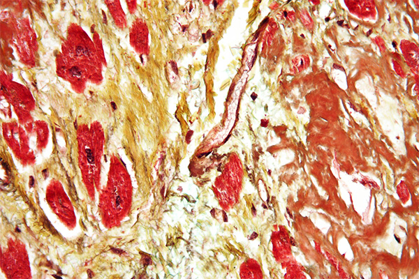
Hart’s Stain for Elastic Fibres

By Dye Type, Elastic Fibers, Iron Resorcin, Protocols, Stain Target, Stain Type

Hart's Stain

for Elastic Fibres

7
steps
4
materials

Materials

Tissue Sample

5µ paraffin sections of neutral buffered formalin fixed tissue are suitable. Other fixatives are likely to be satisfactory.

Protocol

  1. Bring sections to water via xylene and ethanol.
  2. Place into Hart’s solution overnight.
  3. Wash with 95% ethanol to remove excess solution.
  4. Differentiate with 1% acid alcohol.
  5. Wash with tap water.
  6. Counterstain with iron hematoxylin and van Gieson.
  7. Dehydrate with ethanol, clear with xylene and mount with a resinous medium.

Expected Results

  • Elastic fibres  –  blue black
  • Collagen  –  red
  • Cytoplasm  –  yellow
  • Nuclei  –  blue

Notes

  • This modification should be used if poor staining is obtained using Weigert’s method.
  • The optimum dilution may be more or less than given.

Safety Note

Prior to handling any chemical, consult the Safety Data Sheet (SDS) for proper handling and safety precautions.

References

  1. Culling C.F.A., (1974)
    Handbook of histopathological and histochemical techniques Ed. 3
    Butterworth, London, UK.

Fullmer & Lillie’s Orcinol-New Fuchsin for Elastic Fibres

By Dye Type, Elastic Fibers, Iron Resorcin, Protocols, Stain Target, Stain Type

Fullmer & Lillie's Orcinol-New Fuchsin

for Elastic Fibres

5
steps
7
materials

Materials

    • Weigert’s iron hematoxylin or equivalent
    • Van Gieson’s picro-fuchsin
    • Orcinol-New Fuchsin solution
      MaterialAmount
      New fuchsin2g
      Orcinol4g
      Ferric chloride, 30% aqu.50mL
      Distilled water200mL
      Ethanol 95%100mL

      Preparation

      1. Add the dye and orcinol to the water in an oversize flask.
      2. Boil for 5 minutes.
      3. Add the ferric chloride.
      4. Boil for a further 5 minutes.
      5. Cool and filter.
      6. Dry the filter paper and beaker. Place the precipitate and filter paper back into the flask.
      7. Add the ethanol and heat carefully until the precipitate dissolves.
      8. Restore to 100 mL with 95% ethanol.

Tissue Sample

5µ paraffin sections of neutral buffered formalin fixed tissue are suitable. Other fixatives are likely to be satisfactory.

Protocol

  1. Bring sections to water via xylene and ethanol.
  2. Place into orcinol-new fuchsin solution for 15 minutes at 37°C.
  3. Wash with 3 changes of 70% ethanol for 5 minutes each.
  4. Counterstain with iron hematoxylin and van Gieson.
  5. Dehydrate with ethanol, clear with xylene and mount with a resinous medium.

Expected Results

  • Elastic fibres  –  deep violet
  • Collagen  –  red
  • Cytoplasm  –  yellow
  • Nuclei  –  black

Notes

  • Fullmer and Lillie specified 25mL USP IX Liquor Ferri Chloridi, or a solution of 15.5g ferric chloride (FeCl3.6H2O) made up to 25 mL with water, i.e. 25mL of a 62% solution. In either case, it should be fresh.

Safety Note

Prior to handling any chemical, consult the Safety Data Sheet (SDS) for proper handling and safety precautions.

References

  1. Fullmer, H M and Lillie, R D, (1956).
    A selective stain for elastic tissue (orcinol-new fuchsin).,
    Stain Technologyl, v 31, page 27-29.
  2. Lillie, R D and Fullmer, H M, (1976).
    Histopathological technic and practical histochemistry, Ed. 4. pp. 714.
    McGraw-Hill, New York, USA.

Dahlia-Iron-Resorcin for Elastic Fibres

By Dye Type, Elastic Fibers, Iron Resorcin, Protocols, Stain Target, Stain Type

Dahlia-Iron-Resorcin

for Elastic Fibres

8
steps
9
materials

Materials

    • Weigert’s iron hematoxylin or equivalent
    • Van Gieson’s picro-fuchsin
    • Iron-resorcin-dahlia solution
      MaterialAmount
      Dahlia2g
      Resorcin4g
      Ferric nitrate, 30% aqu.50mL
      0.5% aqu. hydrochloric acid200mL
      Methanol180mL
      Acetone20mL
      Hydrochloric acid, conc.4mL

Preparation

  1. Bring 200 mL of 0.5% hydrochloric acid in an oversize flask to the boiling point, remove from the heat and carefully add the dahlia.
  2. Return to the boil, and add the resorcin and ferric nitrate.
  3. Continue boiling for 3 minutes, then cool and filter. Discard the filtrate. Dry the filter paper and beaker, then place the precipitate and filter paper back into the flask.
  4. Add the methanol, acetone and hydrochloric acid.
  5. Heat carefully on a hot plate until the precipitate dissolves.

Tissue Sample

5µ paraffin sections of neutral buffered formalin fixed tissue are suitable. Other fixatives are likely to be satisfactory.

Protocol

  1. Bring sections to water via xylene and ethanol.
  2. Do a Mallory bleach.
  3. Place into the staining solution for 30 minutes or longer.
  4. Wash with 95% ethanol to remove excess solution.
  5. Differentiate with 1% acid alcohol if necessary.
  6. Wash in water.
  7. Counterstain with iron hematoxylin and van Gieson.
  8. Dehydrate with ethanol, clear with xylene and mount with a resinous medium.

Expected Results

  • Elastic fibres  –  blue black
  • Collagen  –  red
  • Cytoplasm  –  yellow
  • Nuclei  –  blue

Notes

  • Between the 1st and 2nd editions of their book, Bancroft and Stevens changed the solvent for the precipitate to the following:
    SolventAmount
    2-methoxyethanol100mL
    Distilled water100mL
    Hydrochloric acid, conc.4mL

    Dry the filter paper and beaker, then place the precipitate and filter paper back into the flask. Add the mixture and heat carefully on a hot plate until the precipitate dissolves.

  • 2-methoxyethanol is also known as ethylene glycol monomethyl ether.
  • The authors suggest the following dyes as suitable substitutes for dahlia:
    • Methyl violet
    • Ethyl violet
    • Victoria blue
    • Thionin
  • Note that this method uses ferric nitrate instead of ferric chloride.

Safety Note

Prior to handling any chemical, consult the Safety Data Sheet (SDS) for proper handling and safety precautions.

References

  1. Bancroft, J. D. and Stevens, A.,
    Theory and Practice of Histological Techniques, Ed. 1 (1977),
    Churchill Livingstone, London, UK.
  2. Bancroft, J. D. and Stevens, A.,
    Theory and Practice of Histological Techniques, Ed. 2 (1982),
    Churchill Livingstone, London, UK.

Silverman-Movat Pentachrome for Elastic, Mucin & Collagen

By Dye Type, Elastic Fibers, Orcein, Protocols, Stain Target, Stain Type

Silverman-Movat Pentachrome

for Elastic, Mucin & Collagen

18
steps
18
materials

Expected Results

  • Nuclei  –  dark purple to black
  • Elastic fibres  –  purple to black
  • Muscle  –  Red, with longitudinal myofibrils, cross striations and intercalated discs delineated
  • Collagen and bone  –  yellow to yellow green
  • Mucin  –  blue
  • Ground substance  –  blue green
  • Cytoplasm  –  pink to brownish to red

Materials

Stock solution A

MaterialAmount
Orcein1g
Hydrochloric acid, conc.1mL
Ethanol, 70%500mL

Stock solution B

MaterialAmount
Hematoxylin8g
Ethanol, absolute160mL

Stock solution C

MaterialAmount
Ferric chloride9.6g
Distilled water90mL

Stock solution D

MaterialAmount
Iodine1g
Potassium iodide2g
Distilled water97mL

Working elastic solution

MaterialAmount
Stock solution A25mL
Stock solution B8mL
Stock solution C5mL
Stock solution D5mL

Differentiator

MaterialAmount
Stock C10mL
Distilled water40mL

Alcian blue

MaterialAmount
Alcian blue1g
Acetic acid, glacial1mL
Distilled water99mL

Ammoniated ethanol

MaterialAmount
Strong ammonia5mL
Ethanol, 95%95mL

Plasma stain

MaterialAmount
Woodstain scarlet 1% aqu.4mL
Acid fuchsin 1% aqu.1mL
Acetic acid, 0.5%95mL

Polyacid

MaterialAmount
Phosphotungstic acid5g
Distilled water100mL

Fiber stain

MaterialAmount
Spanish saffron6g
Ethanol, absolute96mL

Preparation

  1. Incubate together in a sealed container at 56°C for 48 hours. Cool.

Tissue Sample

Paraffin sections at 4-6µ are suitable. Bouin’s fixation is preferred. 10% neutral buffered formalin is satisfactory. If formalin is used, refix sections with Bouin’s fluid for one hour at 56°C, then wash in running tap water to remove the yellow colour.

Protocol

  1. Bring sections to water via xylene and ethanol.
  2. Stain in alcian blue for 20 min.
  3. Rinse in distilled water.
  4. Place in ammoniated ethanol for 10 min at 56°C.
  5. Wash in running tap water for 2 min.
  6. Rinse in distilled water.
  7. Stain in working elastic solution for 2 hours.
  8. Wash in running water until collagen is clear and elastic prominent.
  9. Rinse in distilled water.
  10. Place in the plasma stain for 3 minutes.
  11. Place in 0.5% aqueous acetic acid for 30 seconds.
  12. Differentiate in the polyacid until collagen is clear and ground substance is blue.
  13. Rinse in 0.5% aqueous acetic acid for 30 seconds.
  14. Place in three changes of absolute ethanol for 1 minute each.
  15. Place in the fiber stain for 8 minutes.
  16. Dehydrate quickly in absolute ethanol, 2 changes.
  17. Clear in xylene, three changes.
  18. Coverslip with a resinous mounting medium.

Notes

  • The working elastic solution should be used only once, then discarded.
  • If the elastic fibers are not clearly delineated at step 8 and the background is not clean, place in the differentiator for a few minutes, then wash well in tap water until they are sharp.
  • Differentiation with the polyacid at step 12 takes from 3-10 minutes.
  • The fiber stain (alcoholic saffron) may be used repeatedly until staining intensity decreases. It is important that this solution not be contaminated with water. Place some Drierite into it to keep it anhydrous.

Safety Note

Prior to handling any chemical, consult the Safety Data Sheet (SDS) for proper handling and safety precautions.

References

  1. Silverman, J., (1972)
    Histologic v 2, N° 2.

Garvey-Movat Pentachrome for Elastic, Mucin & Collagen

By Dye Type, Elastic Fibers, Iron Hematoxylin, Protocols, Stain Target, Stain Type

Garvey-Movat Pentachrome

for Elastic, Mucin & Collagen

13
steps
17
materials

Expected Results

  • Nuclei  –  black
  • Elastic fibres  –  black
  • Fibrin – mature  –  red
  • Muscle  –  red
  • Collagen  –  yellow
  • Ground substance  –  blue-green

Materials

Stock Verhoeff A

MaterialAmount
Hematoxylin3g
Ethanol, absolute100mL

Stock Verhoeff B

MaterialAmount
Ferric chloride2.3g
Distilled water100mL

Stock Verhoeff C

MaterialAmount
Potassium iodide4g
Iodine2g
Distilled water100mL

Working Verhoeff

MaterialAmount
Stock solution A3volumes
Stock solution B2volumes
Stock solution C1volume

Polyacid

MaterialAmount
Phosphotungstic acid2g
Distilled water100mL

Acetic water

MaterialAmount
Acetic acid, glacial0.5mL
Distilled water100mL

Picro-mercuric chloride

MaterialAmount
Picric acid, sat. aqu.100mL
Mercuric chloride5g
Distilled water100mL

Alcian blue

MaterialAmount
Alcian blue1g
Acetic acid, glacial3mL
Distilled water100mL

Plasma stain

MaterialAmount
Biebrich scarlet, 1% aqu.8mL
Acid fuchsin, 1% aqu.2mL
Acetic acid, 1% aqu.100mL

Fibre stain

MaterialAmount
Saffron du Gatinais6g
Ethanol, absolute100mL

Preparation

  1. Add the saffron to the ethanol and seal the container.
  2. Incubate at 56°C for 2 weeks. This solution must be anhydrous.

Tissue Sample

Formol sublimate fixation is preferred, although 10% formalin variants are acceptable. Paraffin sections at 3µ are preferred.

Protocol

  1. Bring sections to water via xylene and ethanol.
    1. Optionally, place into picro-mercuric chloride for 1 hour.
    2. Wash well with warm water to remove picric acid.
  2. Rinse with 3% acetic acid.
  3. Place in alcian blue at 60°C for 10 minutes
  4. Rinse with distilled water.
  5. Place in working Verhoeff’s solution for 6 minutes.
  6. Wash with warm tap water for 6 minutes.
  7. Place in the plasma stain for 3 minutes
  8. Rinse with distilled water.
  9. Place in the polyacid for 15 minutes.
  10. Rinse with 1% acetic acid.
  11. Dehydrate thoroughly with absolute ethanol 3 changes.
  12. Place in the fiber stain for 5-6 minutes.
  13. Dehydrate, clear and mount with a resinous medium

Notes

  • Sections should be treated with picro-mercuric chloride if formol sublimate or similar fixation was not used initially.
  • The ferric chloride should be the hexahydrate crystals.
  • Sections thicker than 4µ may need the elastic stain differentiated by treating with 1% aqueous ferric chloride for 20-30 seconds, then washing well with tap water.
  • Although the iodine content of the Verhoeff elastic stain component may remove any mercury pigment, in methods such as this, some technologists prefer to apply the iodine-thiosulphate sequence before staining.

Safety Note

Prior to handling any chemical, consult the Safety Data Sheet (SDS) for proper handling and safety precautions.

References

  1. Garvey W., et. al., (1986),
    Improved Movat pentachrome stain
    Stain Technology, V. 61, No 1, pp 60-62.

Garvey’s Stain for Elastic, Fibrin & Collagen

By Dye Type, Elastic Fibers, Fibrin, Iron Hematoxylin, Protocols, Stain Target, Stain Type

Garvey's Stain

for Elastic, Fibrin & Collagen

12
steps
19
materials

Materials

Stock Verhoeff A

MaterialAmount
Hematoxylin3g
Ethanol, absolute100mL

Stock Verhoeff B

MaterialAmount
Ferric chloride2.3g
Distilled water100mL

Stock Verhoeff C

MaterialAmount
Potassium iodide4g
Iodine2g
Distilled water100mL

Working Verhoeff

MaterialAmount
Stock solution A3volumes
Stock solution B2volumes
Stock solution C1volume

Polyacid

MaterialAmount
Phosphotungstic acid2g
Distilled water100mL

Acetic water

MaterialAmount
Acetic acid, glacial0.5mL
Distilled water100mL

Picro-mercuric chloride

MaterialAmount
Picric acid, sat. aqu.100mL
Mercuric chloride5g
Distilled water100mL

Erythrocyte stain

MaterialAmount
Lissamine fast yellow1g
Acetic acid, glacial0.5mL
Distilled water100mL

Plasma stain

MaterialAmount
Biebrich scarlet0.75g
Acid fuchsin0.75g
Ponceau 2R0.75g
Acetic acid, glacial0.5mL
Distilled water100mL
MaterialAmount for Var IAmount for Var II
Aniline blue0.5g
Light green SF yellowish2g
Acetic acid, glacial1mL2mL
Distilled water100mL100mL

Tissue Sample

Formol sublimate fixation is preferred, although 10% formalin variants are acceptable. Paraffin sections at 3µ are preferred.

Protocol

  1. Bring sections to water via xylene and ethanol.
    1. Optionally, place into picro-mercuric chloride for 1 hour.
    2. Wash well with warm water to remove picric acid.
  2. Place in verhoeff’s solution for 9 minutes.
  3. Wash with warm tap water for 5 minutes.
  4. Place in lissamine fast yellow for 2 minutes.
  5. Rinse with 0.5% acetic acid.
  6. Place in the plasma stain for 5 minutes.
  7. Rinse with distilled water.
  8. Place in polyacid for 10 minutes.
  9. Rinse with distilled water.
  10. Place in a fiber stain variant for 2 minutes.
  11. Rinse with 0.5% acetic acid.
  12. Dehydrate, clear and mount with a resinous medium.

Expected Results

  • Nuclei  –  black
  • Elastic fibres  –  black
  • Fibrin – young  –  yellow
  • Fibrin – mature  –  red
  • Fibrin – old  –  blue or green
  • Erythrocytes  –  yellow
  • Muscle  –  red
  • Collagen  –  blue or green (some may be red)

Notes

  • Sections should be treated with picro-mercuric chloride if formol sublimate or similar fixation was not used initially.
  • The ferric chloride should be the hexahydrate crystals.
  • Although the iodine content of the Verhoeff elastic stain component may remove any mercury pigment, in methods such as this, some technologists prefer to apply the iodine-thiosulphate sequence before staining.

Safety Note

Prior to handling any chemical, consult the Safety Data Sheet (SDS) for proper handling and safety precautions.

References

  1. Garvey W., et. al., (1987),
    A combined elastic, fibrin and collagen stain
    Stain Technology, V. 62, No 6, pp 365-367.

Pasini’s Trichrome for Elastic and Collagen

By Dye Type, Elastic Fibers, Orcein, Protocols, Stain Target, Stain Type, Trichrome Staining, Trichrome, One-Step

Pasini's Trichrome

for Elastic and Collagen

8
steps
8
materials

Materials

  • Ehrlich’s alum hematoxylin
  • Solution A
    MaterialAmount
    Phosphotungstic acid2g
    Distilled water100mL
  • Solution B – Var I
    MaterialAmount
    Eosin B0.7g
    Acid fuchsin, sat. aqu.4mL
    Unna’s elastic stain35mL
    Glycerol40mL
    Ethanol, 50%35mL
  • Solution B – Var II
    MaterialAmount
    Eosin B, 2% in 50% ethanol30mL
    Acid fuchsin, sat. aqu.4mL
    Unna’s elastic stain30mL
    Glycerol15mL

Tissue Sample

5µ paraffin sections of neutral buffered formalin fixed tissue are suitable. Other fixatives are likely to be satisfactory. Most trichrome stains benefit from picric acid or mercuric chloride fixation. Formalin fixed tissues may benefit from secondary fixation of sections in Bouin’s fluid.

Protocol

  1. Bring sections to water via xylene and ethanol.
  2. Stain nuclei with an alum hematoxylin nuclear stain.
  3. Place into solution A for 10 minute.
  4. Rinse quickly with distilled water.
  5. Place into solution B for 15-20 minutes.
  6. Rinse quickly with 70% ethanol.
  7. Dehydrate and differentiate with 100% ethanol for a few seconds.
  8. Clear with xylene and mount with a resinous medium.

Expected Results

  • Nuclei  –  blue
  • Elastic fibres  –  purple
  • Erythrocytes  –  orange
  • Collagen  –  blue

Notes

  • Solution B var I was given by Gray, var II was given by Gatenby & Beams.

Safety Note

Prior to handling any chemical, consult the Safety Data Sheet (SDS) for proper handling and safety precautions.

References

  1. Gray, Peter. (1954)
    The Microtomist’s Formulary and Guide.
    Originally published by: The Blakiston Co.
    Republished by: Robert E. Krieger Publishing Co.
    Citing:
    Gatenby, J. B. & Cowdry, E. V., (1928)
    Bolles Lee’s Microtomist’s Vade Mecum, pp. 280
    Blakiston, Philadelphia
  2. Bolles Lee, A.. Edited by Gatenby, J.B. and Beams, H.W., (1950)
    The Microtomist’s Vade-Mecum. 11 ed., pp. 485
    Churchill, London, UK.

Mollier’s trichrome for Elastic and Collagen

By Dye Type, Elastic Fibers, Orcein, Protocols, Stain Target, Stain Type, Trichrome Staining, Trichrome, Multi-Step

Mollier's trichrome

for Elastic and Collagen

13
steps
13
materials

Materials

  • Weigert’s iron hematoxylin or equivalent
  • Solution A
    MaterialAmount
    Orcein0.8g
    Hydrochloric acid1mL
    Ethanol, 100%50mL
    Distilled water50mL
  • Solution B
    MaterialAmount
    Azocarmine2g
    Acetic acid, glacial1mL
    Distilled water100mL
  • Solution C
    MaterialAmount
    Phosphotungstic acid5g
    Distilled water100mL
  • Solution D
    MaterialAmount
    Naphthol green B1g
    Acetic acid, glacial1mL
    Distilled water100mL

Tissue Sample

5µ paraffin sections of neutral buffered formalin fixed tissue are suitable. Other fixatives are likely to be satisfactory. Most trichrome stains benefit from picric acid or mercuric chloride fixation. Formalin fixed tissues may benefit from secondary fixation of sections in Bouin’s fluid.

Protocol

  1. Bring sections to water via xylene and ethanol.
  2. Place into solution A for 12 hours.
  3. Stain nuclei with Weigert’s iron hematoxylin for 1-3 minutes.
  4. Differentiate the nuclear stain if necessary.
  5. Wash with water for 15 minutes.
  6. Place into solution B for 15-30 minutes.
  7. Rinse with distilled water.
  8. decolorise in solution C for 2-6 hours, changing the solution three times.
  9. Rinse quickly with distilled water.
  10. Place into solution D for 15-30 minutes.
  11. Agitate vigorously in 95% ethanol for 30 seconds.
  12. Dehydrate with ethanol.
  13. Clear with xylene and mount with a resinous medium.

Expected Results

  • Nuclei  –  blue
  • Elastic  –  black
  • Erythrocytes  –  red
  • Cytoplasm  –  purple
  • Collagen  –  green

Notes

  • Solution A is the Unna-Taenzer elastic solution.

Safety Note

Prior to handling any chemical, consult the Safety Data Sheet (SDS) for proper handling and safety precautions.

References

  1. Gray, Peter. (1954)
    The Microtomist’s Formulary and Guide.
    Originally published by: The Blakiston Co.
    Republished by: Robert E. Krieger Publishing Co.
    Citing:
    Mollier, (1938)
    Zeitschrift für wissenschaftliche Mikroskopie und für mikroskopische Technik, v.55, pp.472.
    And:
    von Kahlden, C. and Laurent, O., (1896)
    Technique microscopique, pp.143
    Carré, Paris, France

Kohashi’s Trichrome for Elastic and Collagen

By Dye Type, Elastic Fibers, Orcein, Protocols, Stain Target, Stain Type, Trichrome Staining, Trichrome, Multi-Step

Kohashi's Trichrome

for Elastic and Collagen

12
steps
14
materials

Materials

Solution A

MaterialAmount
Azocarmine0.1g
Acetic acid, glacial1mL
Distilled water99mL

Solution B

MaterialAmount
Aniline0.1mL
Ethanol, 95%100mL

Solution C

MaterialAmount
Acetic acid, glacial1mL
Ethanol, 95%100mL

Solution D

MaterialAmount
Phosphotungstic acid5g
Distilled water100mL

Solution E

MaterialAmount
Eosin B0.7g
Acid fuchsin, sat. aqu.4mL
Unna’s elastic stain35mL
Ethanol, 50%35mL
Glycerol40mL

Tissue Sample

Many fixatives are satisfactory. 5µ paraffin sections of neutral buffered formalin fixed tissue are suitable. Most trichrome stains benefit from picric acid or mercuric chloride fixation. Formalin fixed tissues may benefit from secondary fixation of sections in Bouin’s fluid.

Protocol

  1. Bring sections to water via xylene and ethanol.
  2. Place into solution A for 12-15 minutes.
  3. Rinse quickly with distilled water.
  4. Differentiate nuclei with solution B.
  5. Place into solution C for 30-60 seconds.
  6. Rinse with distilled water.
  7. Place into solution D for 30-60 minutes.
  8. Rinse with distilled water.
  9. Place into solution E for 15-20 minutes.
  10. Place into 95% ethanol until differentiated.
  11. Dehydrate with ethanol.
  12. Clear with xylene and mount with a resinous medium.

Expected Results

  • Nuclei  –  red
  • Erythrocytes  –  orange
  • Elastic fibres  –  purple
  • Cytoplasm  –  red
  • Collagen  –  blue

Notes

Safety Note

Prior to handling any chemical, consult the Safety Data Sheet (SDS) for proper handling and safety precautions.

References

  1. Gray, Peter. (1954)
    The Microtomist’s Formulary and Guide.
    Originally published by: The Blakiston Co.
    Republished by: Robert E. Krieger Publishing Co.
    Citing:
    Kohashi, (1937)
    Folia anatomica Japonica, v.15, pp.175