Category

Periodic Acid-Schiff Reaction

Periodic Acid Schiff Diastase

By Aldehydes, Periodic Acid-Schiff Reaction, Protocols, Schiff's Reagent Reactions, Stain Target, Stain Type
Protocol

Periodic Acid Schiff Diastase

15
steps
4
materials

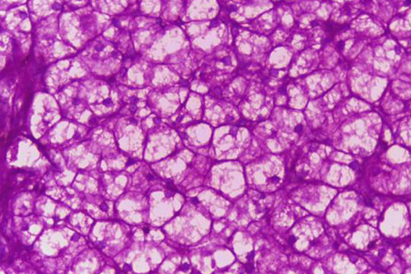
The periodic acid Schiff reaction (PAS) is used to demonstrate the presence of 1-2-glycols, including glycogen. Sometimes it is necessary for the glycogen to be removed so that a clearer picture of non-glycogen carbohydrates may be seen. Glycogen may be removed with amylase (diastase).

Materials

Tissue Sample

5µ paraffin sections of neutral buffered formalin fixed tissue are suitable. Other fixatives are usually satisfactory, although glutaraldehyde should be avoided.

Protocol

  1. Two consecutive test and 2 consecutive control sections.
  2. Bring sections to water via xylene and ethanol.
  3. Place one test section and one known positive section into water.
  4. Place the other test section and the other known positive in the amylase solution for 30-60 minutes at 35-45°C
  5. Wash both digested sections with tap water
  6. Celloidinise the sections if desired.
  7. Place into periodic acid for 10-30 minutes.
  8. Rinse well with tap water.
  9. Rinse with distilled water.
  10. Place in Schiff’s reagent for 10-30 minutes.
  11. Wash off with distilled water.
  12. Wash well with tap water for about 10 minutes.
  13. Counterstain with Mayer’s hemalum for 2 minutes.
  14. Wash well with tap water until hemalum is blued.
  15. Dehydrate with ethanol, clear with xylene and coverslip using a resinous medium.

Expected Results

  • Glycogen – red in the undigested section, absent from the digested section.
  • Oxidisable carbohydrates – red
  • Nuclei – blue

Notes

  • If the intent is to remove glycogen rather than identify it, a more convenient approach in a routine service laboratory is to dissolve the amount of α-amylase that would cover a 1 cm circle about 1 mm deep in 15 mL (1 test tube) of distilled water. Shake for a few minutes, then filter onto the sections and digest at room temperature. Glycogen is usually removed within 30 minutes to 1 hour. Wash well and continue with a regular PAS (step 7).
  • Please note that an older variation of the procedure in Note 1 was to collect saliva and use that for the digestion. This is strongly deprecated. Human body fluids may contain transmissible bacterial and viral contaminants, so anyone handling such a slide will be at risk.
  • Hog α-amylase is recommended as being consistent and effective. Purchase a product with a high α-amylase activity.
  • Glutaraldehyde fixation leaves free aldehyde groups attached to tissues, which causes an overall positive reaction. These groups may be stopped from reacting with an appropriate procedure such as the aniline-acetic aldehyde block.
  • The tap water wash at step 7 is necessary to develop the red color. Within limits, the longer the wash the darker the color.
  • Originally, it was recommended that the Schiff’s reagent be washed off with dilute sulfurous acid (the sulfite rinses). Since water recolors Schiff’s reagent, it was believed that a water wash could lead to false positive results. It is now known this is not the case, provided the Schiff’s reagent is removed quickly and the sections do not stay in water contaminated with it for extended periods.

Safety Note

Prior to handling any chemical, consult the Safety Data Sheet (SDS) for proper handling and safety precautions.

Hotchkiss’ Alcoholic Periodic Acid Schiff

By Aldehydes, Periodic Acid-Schiff Reaction, Protocols, Schiff's Reagent Reactions, Stain Target, Stain Type
Protocol

Hotchkiss' Alcoholic Periodic Acid Schiff

13
steps
10
materials

It was initially thought that the periodic acid Schiff reaction could result in less glycogen being demonstrated than was actually present because it might dissolve in aqueous reagents. It is now known this is not a concern. Hotchkiss recommended an alcoholic method to ensure it did not take place. This method is now redundant.

Materials

  • Schiff’s reagent
  • Mayer’s hemalum
  • Alcoholic periodic acid
    MaterialAmount
    Periodic acid0.8g
    Sodium acetate buffer 0.2M10mL
    Ethanol, absolute70mL
    Distilled water20mL
  • Acid reducing rinse
    MaterialAmount
    Potassium iodide2g
    Sodium thiosulphate2g
    Ethanol absolute60mL
    Hydrochloric acid N/12mL
    Distilled water40mL

Tissue Sample

Presumably an alcoholic fixative should be required if glycogen were to dissolve in aqueous solutions. However, 5 µ paraffin sections of neutral buffered formalin fixed tissue are suitable. Other fixatives are usually satisfactory, although glutaraldehyde should be avoided.

Protocol

  1. Bring sections to water via xylene and ethanol.
  2. Place into periodic acid for 10 minutes.
  3. Rinse with 70% ethanol.
  4. Place in acid reducing rinse for 1 minute.
  5. Rinse with 70% ethanol.
  6. Wash with running water to remove ethanol.
  7. Rinse with distilled water.
  8. Place in Schiff’s reagent for 10-30 minutes.
  9. Wash off with distilled water.
  10. Wash well with tap water for about 10 minutes.
  11. Counterstain with Mayer’s hemalum for 1 minutes.
  12. Wash well with tap water until hemalum is blued.
  13. Dehydrate with ethanol, clear with xylene, and coverslip using a resinous medium.

Expected Results

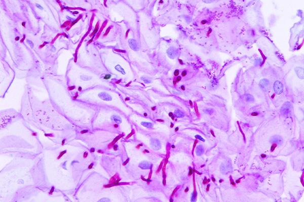
  • Oxidisable carbohydrates – red
  • Nuclei – blue

Safety Note

Prior to handling any chemical, consult the Safety Data Sheet (SDS) for proper handling and safety precautions.
References

  1. Culling C.F.A., (1963)
    Handbook of histopathological and histochemical techniques Ed. 2
    Butterworth, London, UK.

Double Oxidation Schiff

By Aldehydes, Periodic Acid-Schiff Reaction, Protocols, Schiff's Reagent Reactions, Stain Target, Stain Type

Double Oxidation Schiff

14
steps
3
materials

Double oxidation may be used with any procedure which demonstrates aldehydes, including Schiff’s reagent and methenamine silver reduction. Its use is not necessarily confined to staining fungi and it may be found useful when demonstrating other structures and the background stains too darkly.

Materials

  • Periodic acid – 1% aqueous
  • Analine-acetic block
    MaterialAmount
    Acetic acid, glacial5mL
    Aniline oil45mL

Tissue Sample

5µ paraffin sections of neutral buffered formalin fixed tissue are suitable.

Protocol

  1. Bring sections to water via xylene and ethanol.
  2. Oxidize with periodic acid for 10 minutes.
  3. Rinse well with tap water.
  4. Treat with aniline-acetic for 30 minutes.
  5. Rinse well with tap water.
  6. Re-oxidize with periodic acid for 20 minutes.
  7. Rinse well with tap water.
  8. Rinse with distilled water.
  9. Place in Schiff’s reagent for 10-30 minutes.
  10. Wash off with distilled water.
  11. Wash well with tap water for about 10 minutes.
  12. Counterstain with Mayer’s hemalum for 2 minutes.
  13. Wash well with tap water until hemalum is blued.
  14. Dehydrate with ethanol, clear with xylene and coverslip using a resinous medium.

Expected Results

  • Oxidizable carbohydrates  –  red
  • Fungi  –  red
  • Nuclei & background  –  blue

Notes

  • The time periodic acid is applied in the first oxidation determines the amount of background staining eliminated. If there is only a small amount of this material, then all of it may be blocked
  • The first oxidation will also oxidize some of the target material, and this staining will also be inhibited. If the first oxidation is applied for too long, the depth of staining of the target material may be affected. The 10 minutes specified is usually enough.
  • The time periodic acid is applied in the second oxidation determines the depth of staining for the target material. 20 minutes is usually adequate, but extending it may give darker staining. At some point, extending the time in periodic acid will cease to produce darker staining as all available carbohydrate will have been oxidized.

Safety Note

Prior to handling any chemical, consult the Safety Data Sheet (SDS) for proper handling and safety precautions.

References

  1. Personal observation, B. Llewellyn,

Lillie’s Long PAS for 1-2-Glycols

By Aldehydes, Periodic Acid-Schiff Reaction, Protocols, Schiff's Reagent Reactions, Stain Target, Stain Type

Lillie's Long PAS

for 1-2-Glycols

14
steps
6
materials

The periodic acid Schiff reaction (PAS) is used to demonstrate the presence of 1-2-glycols, and is consequently an important method in the histochemistry of carbohydrates and the histological demonstration of many structures.

Lillie referred to this as

“The Periodic acid Leucofuchsin Method in Relation to Various Modifying Procedures”.

He describes its purpose as being

“To indicate at what points and in what sequence various modifying procedures should be introduced”.

Materials

  • A periodic acid solution.
  • A strong Schiff’s reagent.
  • Sodium metabisulfite, 0.5% aqueous, optional.
  • Mayer’s hemalum, or Weigert’s iron hematoxylin.
  • Picric acid, saturated aqueous, optional.
  • Orange G, 2% aqueous, optional.
  • Methyl blue, 0.1% in saturated aqueous picric acid, optional.

Tissue Sample

5µ paraffin sections of neutral buffered formalin fixed tissue are suitable. Other fixatives are likely to be satisfactory, although glutaraldehyde should be avoided.

Protocol

  1. Dewax paraffin sections with xylene.
  2. Remove xylene with two changes of absolute ethanol.
  3. Optionally, do one or more of the following procedures in the order given.
    ProcedureComments
    AcetylationAcetylation blocks hydroxyl and amino groups. Cartilage and glycogen are the most resistant. Other carbohydrates are more easily affected.
    BenzoylationSimilar to acetylation, this also blocks hydroxyl and amino groups. Again, cartilage and glycogen are the most resistant while other carbohydrates are more easily affected.
    Deacetylation (saponification)Deacetylation (also called saponification) reverses the effects of acetylation in some, if not all of the materials which were blocked. The reversal is progressive, that is, some substances recover their stainabilty before others.
    MethylationExtended methylation causes some otherwise oxidisable carbohydrates to become non-reactive. Primarily, however, it inhibits basophilia.
    Aldehyde blockIf applied before periodic acid oxidation, pre-existing aldehydes are inhibited from reacting and will be unstained. If applied after periodic acid oxidation, aldehydes produced by the periodic acid are inhibited from reacting and will be unstained. A second section which has not been blocked can be used to prove that oxidation formed the aldehydes.
    Amylase digestionAmylase (diastase) is used to remove glycogen. This is either to prove that a subtance is glycogen, or to remove it so that other PAS positive materials other than glycogen may be more clearly seen.
    Protease digestionProteases may also be used to remove carbohydrate-protein complexes, although this is less common than amylase digestion because the enzymes also digest other tissue proteins.
    Reducing rinsesThis is an acidified solution of potassium iodide and sodium thiosulphate applied following periodic acid to remove any iodate or periodate remaining in the tissue.
    Methanol, Pyridine & Solvent extractionPAS positive carbohydrate-lipid complexes, or glycolipids, may be removed with these solvents. They may be used at ambient or elevated temperature.
  4. Ensure sections are in water or an appropriate concentration of ethanol.
  5. Oxidize for the appropriate time in the selected periodic acid solution.
  6. Wash five minutes in running water, or 70% ethanol if using an alcoholic variant.
  7. Optionally, do one of the following.
    1. Hotchkiss’ reducing rinse, or
    2. Aldehyde block
  8. Place in Schiff’s reagent for 10 minutes, or longer if specially required.
  9. Transfer to three successive sulfite rinses, 2 minutes each.
  10. Wash in running tap water for 10 minutes.
  11. Optionally, stain nuclei.
  12. Optionally, stain cytoplasm.
  13. Dehydrate and differentiate with two changes each of 95% and absolute ethanol.
  14. Clear with xylene and coverslip using a resinous medium.

Expected Results

  • 1-2-glycols  –  red
  • Nuclei  –  blue or black
  • Cytoplasm  –  yellow or unstained

Notes

  • A strong Schiff’s reagent is recommended, i.e. one that contains 0.5 or 1 gram of pararosanilin per 200 mL solution.
  • Sections should be celloidinised before deacetylation, and it may be advisable after the other options in step 3.
  • Experience shows that the reducing rinses may be omitted.
  • Mayer’s hemalum or Weigert’s iron hematoxylin are suitable nuclear stains.
  • Sat. picric acid, 2% orange G, or picro-methyl blue are suitable cytoplasmic stains.

Safety Note

Prior to handling any chemical, consult the Safety Data Sheet (SDS) for proper handling and safety precautions.

References

  1. Lillie, R.D., (1954)
    Histopathologic technique and practical histochemistry Ed.2
    Blakiston, New York, USA.

Lillie’s Short PAS Reaction for 1-2 Glycols

By Aldehydes, Periodic Acid-Schiff Reaction, Protocols, Schiff's Reagent Reactions, Stain Target, Stain Type

Lillie's Short PAS Reaction

for 1-2 Glycols

8
steps
5
materials

The periodic acid Schiff reaction (PAS) is used to demonstrate the presence of 1-2-glycols, and is consequently an important method in the histochemistry of carbohydrates and the histological demonstration of many structures.

Lillie referred to this method as

The Periodic acid, Schiff Sulfite Leucofuchsin Reaction, short variant.

Materials

Tissue Sample

5µ paraffin sections of neutral buffered formalin fixed tissue are suitable. Other fixatives are likely to be satisfactory, although glutaraldehyde should be avoided.

Protocol

  1. Bring sections to water via xylene and ethanol.
  2. Oxidize in periodic acid for ten minutes.
  3. Wash in running water for five minutes.
  4. Place in Schiff’s reagent for 10 minutes.
  5. Transfer to three successive baths of sodium metabisulfite for 1, 2, and 2 minutes respectively.
  6. Wash in running tap water for 5 minutes.
  7. Counterstain with one of the following:
    1. Place in Mayer’s hemalum for 2 minutes. Wash in running tap water and blue.
    2. Place in Weigert’s iron hematoxylin for 2-4 minutes. Decolorise with Pal’s bleach diluted 1:5 with distilled water. Wash in running tap water for 4 minutes.
    3. Place in Weigert’s iron hematoxylin for 6 minutes. Wash for 4 minutes in running tap water. Place in saturated aqueous picric acid for 1 minute.
  8. Dehydrate with ethanol, clear with xylene, and coverslip using a resinous medium.

Expected Results

  • 1-2-glycols  –  red
  • Nuclei  –  blue or black
  • Cytoplasm  –  yellow or unstained

Notes

  • The nuclear counterstain may obscure some positive staining. Keep the application time short.
  • Weigert’s solution should be allowed to ripen for an hour before use.
  • Weigert’s hematoxylin and Pal’s bleach combination gives highly selective nuclear staining.
  • Picric acid differentiates the nuclear stain as well as coloring the background.
  • Glutaraldehyde fixed tissues will have a non-specific positive background staining. This must be blocked before step 2.

Safety Note

Prior to handling any chemical, consult the Safety Data Sheet (SDS) for proper handling and safety precautions.
References

  1. Lillie, R.D., (1954)
    Histopathologic technique and practical histochemistry Ed.2
    Blakiston, New York, USA.

McManus’ PAS Reaction for 1-2 Glycols

By Aldehydes, Periodic Acid-Schiff Reaction, Protocols, Schiff's Reagent Reactions, Stain Target, Stain Type

McManus’ PAS Reaction

for 1-2 Glycols

8
steps
5
materials

The periodic acid Schiff reaction (PAS) is used to demonstrate the presence of 1-2-glycols, and is consequently an important method in the histochemistry of carbohydrates and the histological demonstration of many structures.

Materials

  • Periodic acid (0.5% aqueous specified).
  • Schiff’s reagent (Coleman’s specified).
  • Harris’ hemalum
  • Light green working solution (0.2% aqueous Light Green diluted 1:5 with distilled water)
  • Ammonia water (water with 3 drops concentrated ammonia per 100 mL)

Tissue Sample

6 µ paraffin sections of neutral buffered formalin fixed tissue are suitable. Other fixatives are likely to be satisfactory, although glutaraldehyde should be avoided.

Protocol

  1. Bring sections to distilled water via xylene and ethanol.
  2. Digest using a diastase, hyaluronidase, or sialidase procedure.
  3. Oxidize in Periodic Acid for 5 minutes.
  4. Rinse in distilled water.
  5. Place in Coleman’s or another Schiff’s Reagent for 15 minutes.
  6. Wash in running water for 10 minutes to develop the pink color.
  7. Counterstain with one of the following:–
    1. Harris’ hematoxylin for 6 minutes, then
      1. Wash in running water and transfer to 1% acid ethanol for 3-10 quick dips
      2. Transfer to 1% acid ethanol for 3-10 quick dips
      3. Wash in distilled water
      4. Dip in ammonia water to blue the sections
      5. Wash in running water for ten minutes
    2. Light green working solution for 10 seconds.
  8. Dehydrate with ethanol, clear with xylene, and coverslip using a resinous medium.

Expected Results

  • 1-2-glycols  –  red
  • Nuclei  –  blue
  • Background  –  green (if light green used)

Notes

  • Light Green is better used when delineation of fungi is required.
  • Tap water and ammonia decolorize Light Green, so proceed directly to dehydration.
  • Glutaraldehyde fixed tissues will have a non-specific positive background staining. This must be blocked before step 2.

Safety Note

Prior to handling any chemical, consult the Safety Data Sheet (SDS) for proper handling and safety precautions.

References

  1. McManus, J. F. A., (1946)
    Stain Technology, v23, p99.

Periodic Acid Schiff Reaction

By Aldehydes, Periodic Acid-Schiff Reaction, Protocols, Schiff's Reagent Reactions, Stain Target, Stain Type

Periodic Acid Schiff Reaction

10
steps
3
materials

The periodic acid Schiff reaction (PAS) is used to demonstrate the presence of 1-2-glycols, and is consequently an important method in the histochemistry of carbohydrates and the histological demonstration of many structures.

Expected Results

  • 1-2-glycols  –  red or dark purple
  • Nuclei  –  blue

Materials

Tissue Sample

5µ paraffin sections of neutral buffered formalin fixed tissue are suitable. Other fixatives are usually satisfactory, although glutaraldehyde should be avoided.

Protocol

  1. Bring sections to water via xylene and ethanol.
  2. Place into periodic acid for 10-30 minutes.
  3. Rinse well with tap water.
  4. Rinse with distilled water.
  5. Place in Schiff’s reagent for 10-30 minutes.
  6. Wash off with distilled water.
  7. Wash well with tap water for about 10 minutes.
  8. Counterstain with Mayer’s hemalum for 2 minutes.
  9. Wash well with tap water until hemalum is blued.
  10. Dehydrate with ethanol, clear with xylene and coverslip using a resinous medium.

Notes

  • Glutaraldehyde fixation leaves free aldehyde groups attached to tissues, which causes an overall positive reaction. These groups may be stopped from reacting with an appropriate procedure such as the aniline-acetic aldehyde block.
  • The tap water wash at step 7 is necessary to develop the red color. Within limits, the longer the wash the darker the color.
  • Originally, it was recommended that the Schiff’s reagent be washed off with dilute sulfurous acid (the sulfite rinses). Since water recolors Schiff’s reagent, it was believed that a water wash could lead to false positive results. It is now known this is not the case, provided the Schiff’s reagent is removed quickly and the sections do not stay in water contaminated with it for extended periods.

Safety Note

Prior to handling any chemical, consult the Safety Data Sheet (SDS) for proper handling and safety precautions.

References

  1. Culling C.F.A., (1974)
    Handbook of histopathological and histochemical techniques Ed. 3
    Butterworth, London, UK.

Lillie’s Allochrome

By Periodic Acid-Schiff Reaction, Protocols, Schiff's Reagent Reactions, Stain Type
Protocol

Lillie's Allochrome

8
steps
5
materials

Materials

  • 1% Aqueous periodic acid
  • Schiff’s reagent
  • Weigert’s iron hematoxylin
  • Picro-methyl blue
    MaterialAmount
    Saturated aqueous picric acid100mL
    1% aqueous methyl blue0.4mL

Tissue Sample

5 µ paraffin sections. Most fixatives are satisfactory, including 10% NBF, but those generally considered preferable for trichrome stains will be advantageous.

Protocol

  1. Bring sections to water via xylene and ethanol.
  2. Do a PAS stain, but do not counterstain.
  3. Stain nuclei with Weigert’s iron hematoxylin or equivalent.
  4. Wash well in tap water, rinse with distilled water.
  5. Place into picro-methyl blue for 6 minutes.
  6. Rinse well with 95% ethanol.
  7. Dehydrate with absolute ethanol.
  8. Clear with xylene and mount with a resinous medium.

Expected Results

  • Nuclei – Brown-black
  • Collagen & reticulin – blue
  • Cytoplasm & muscle – yellow
  • PAS positive structures – red

Safety Note

Prior to handling any chemical, consult the Safety Data Sheet (SDS) for proper handling and safety precautions.

References

  1. Histological demonstration techniques, (1974)
    Cook, H C.
    Butterworths, London, England

Periodic Acid Fluorescent Schiff

By Aldehydes, Periodic Acid-Schiff Reaction, Protocols, Schiff's Reagent Reactions, Stain Target, Stain Type

Periodic Acid Fluorescent Schiff

9
steps
2
materials

Materials

Tissue Sample

5µ paraffin sections of neutral buffered formalin fixed tissue are suitable. Other fixatives are likely to be satisfactory.

Protocol

  1. Dewax and hydrate sections.
  2. Place in periodic acid for 10 minutes.
  3. Wash well with tap water.
  4. Rinse with distilled water.
  5. Place in fluorescent Schiff reagent for 20 minutes.
  6. Wash well with tap water.
  7. Place in acid alcohol, two changes, 5 minutes each.
  8. Wash well with tap water.
  9. Dehydrate, clear and mount in a non fluorescent resinous medium.

Expected Results

Using a BG 12 exciter filter, and OG 4 (yellow) and/or OG5 (orange) barrier filter, 1-2-glycols fluoresce yellow with acriflavine and green-red with acridine orange.

Periodic acid fluorescent schiff

Notes

  • The same materials fluoresce as would be red or pink in a regular PAS reaction.
  • If background fluorescence is too bright it may be quenched with alum hematoxylin for 1 minute,or 0.5% aqueous potassium permanganate for 1 minute. Quench immediately before the final dehydration step.This should be done with caution as it may reduce fluorescence.

Safety Note

Prior to handling any chemical, consult the Safety Data Sheet (SDS) for proper handling and safety precautions.

References

  1. Culling C.F.A., (1974)
    Handbook of histopathological and histochemical techniques Ed. 3
    Butterworth, London, UK.