Category

Protocols

[facetwp template="protocol_by_facet"]

Paquin & Goddard’s Trichrome for Elastic and Collagen

By Protocols, Stain Type, Trichrome Staining, Trichrome, Multi-Step

Paquin & Goddard's Trichrome

for Elastic and Collagen

13
steps
13
materials

Materials

  • Paquin & Goddard’s iron hematoxylin or equivalent
  • Solution A
    MaterialAmount
    Eosin Y0.7g
    Phloxine0.3g
    Orange G0.1g
    Phosphotungstic acid1g
    Distilled water1L
  • Solution B
    MaterialAmount
    Phosphotungstic acid2g
    Distilled water1L
  • Solution C
    MaterialAmount
    Acetic acid, glacial4mL
    Distilled water1L
  • Solution D
    MaterialAmount
    Aniline blue0.4g
    Acetic acid, glacial10mL
    Distilled water990mL

Tissue Sample

5µ paraffin sections of Masson’s picric-chrome-formalin fixation was specified. Most trichrome stains benefit from picric acid or mercuric chloride fixation. The specified fixation may be compensated for by secondary fixation of sections in Bouin’s fluid.

Masson’s picric-chrome-formalin contains:

MaterialAmount
Picric acid, sat. aqu.187.5mL
Chromium aluminum sulphate7.5g
Formalin, Conc. 40%67.5mL
  1. Add the alum to the formalin and leave for 1 hour.
  2. Add the picric acid solution.
  3. Let stand for 24 hours, and filter.
  4. Return to tissue sample.

Protocol

  1. Bring sections to water via xylene and ethanol.
  2. Stain with Paquin & Goddard’s iron hematoxylin.
  3. Wash with water.
  4. Place into solution A for 5 minutes.
  5. Place into solution B for 5 minutes.
  6. Rinse twice with solution C.
  7. Place into solution D for 5 minutes.
  8. Rinse twice with solution C.
  9. Place into solution B for 5 minutes.
  10. Place into solution C for 30 minutes.
  11. Rinse quickly with 95% ethanol.
  12. Dehydrate with iso-amyl alcohol.
  13. Clear with toluene and mount with a resinous medium.

Expected Results

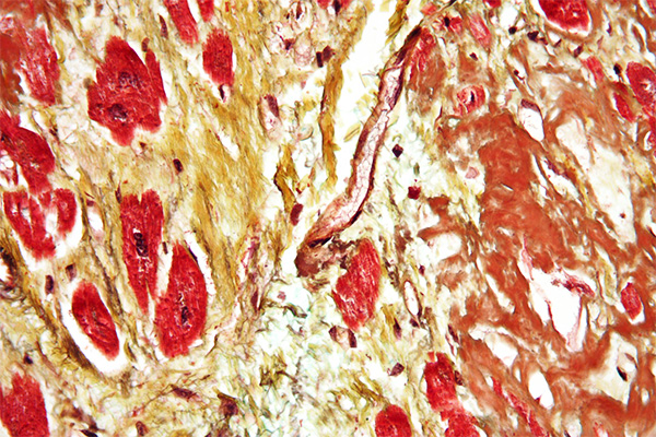
  • Nuclei – black
  • Elastic – red
  • Cytoplasm – pink
  • Collagen – blue

Notes

  • If iso-amyl alcohol is not available, t-butanol is often suitable when the staining is likely to be removed with ethanol, although rapid dehydration with ethanol can often overcome this.
  • Although toluene was specified for clearing, xylene is likely suitable.

Safety Note

Prior to handling any chemical, consult the Safety Data Sheet (SDS) for proper handling and safety precautions.

References

  1. Gray, Peter. (1954)
    The Microtomist’s Formulary and Guide.
    Originally published by: The Blakiston Co.
    Republished by: Robert E. Krieger Publishing Co.
    Citing:
    Paquin & Goddard, (1947)
    Bulletin of the International Association of Medical Museums and
    Journal of Technical Methods, v.27, pp.198

Pasini’s Trichrome for Elastic and Collagen

By Dye Type, Elastic Fibers, Orcein, Protocols, Stain Target, Stain Type, Trichrome Staining, Trichrome, One-Step

Pasini's Trichrome

for Elastic and Collagen

8
steps
8
materials

Materials

  • Ehrlich’s alum hematoxylin
  • Solution A
    MaterialAmount
    Phosphotungstic acid2g
    Distilled water100mL
  • Solution B – Var I
    MaterialAmount
    Eosin B0.7g
    Acid fuchsin, sat. aqu.4mL
    Unna’s elastic stain35mL
    Glycerol40mL
    Ethanol, 50%35mL
  • Solution B – Var II
    MaterialAmount
    Eosin B, 2% in 50% ethanol30mL
    Acid fuchsin, sat. aqu.4mL
    Unna’s elastic stain30mL
    Glycerol15mL

Tissue Sample

5µ paraffin sections of neutral buffered formalin fixed tissue are suitable. Other fixatives are likely to be satisfactory. Most trichrome stains benefit from picric acid or mercuric chloride fixation. Formalin fixed tissues may benefit from secondary fixation of sections in Bouin’s fluid.

Protocol

  1. Bring sections to water via xylene and ethanol.
  2. Stain nuclei with an alum hematoxylin nuclear stain.
  3. Place into solution A for 10 minute.
  4. Rinse quickly with distilled water.
  5. Place into solution B for 15-20 minutes.
  6. Rinse quickly with 70% ethanol.
  7. Dehydrate and differentiate with 100% ethanol for a few seconds.
  8. Clear with xylene and mount with a resinous medium.

Expected Results

  • Nuclei  –  blue
  • Elastic fibres  –  purple
  • Erythrocytes  –  orange
  • Collagen  –  blue

Notes

  • Solution B var I was given by Gray, var II was given by Gatenby & Beams.

Safety Note

Prior to handling any chemical, consult the Safety Data Sheet (SDS) for proper handling and safety precautions.

References

  1. Gray, Peter. (1954)
    The Microtomist’s Formulary and Guide.
    Originally published by: The Blakiston Co.
    Republished by: Robert E. Krieger Publishing Co.
    Citing:
    Gatenby, J. B. & Cowdry, E. V., (1928)
    Bolles Lee’s Microtomist’s Vade Mecum, pp. 280
    Blakiston, Philadelphia
  2. Bolles Lee, A.. Edited by Gatenby, J.B. and Beams, H.W., (1950)
    The Microtomist’s Vade-Mecum. 11 ed., pp. 485
    Churchill, London, UK.

Garvey’s Stain for Elastic, Fibrin & Collagen

By Dye Type, Elastic Fibers, Fibrin, Iron Hematoxylin, Protocols, Stain Target, Stain Type

Garvey's Stain

for Elastic, Fibrin & Collagen

12
steps
19
materials

Materials

Stock Verhoeff A

MaterialAmount
Hematoxylin3g
Ethanol, absolute100mL

Stock Verhoeff B

MaterialAmount
Ferric chloride2.3g
Distilled water100mL

Stock Verhoeff C

MaterialAmount
Potassium iodide4g
Iodine2g
Distilled water100mL

Working Verhoeff

MaterialAmount
Stock solution A3volumes
Stock solution B2volumes
Stock solution C1volume

Polyacid

MaterialAmount
Phosphotungstic acid2g
Distilled water100mL

Acetic water

MaterialAmount
Acetic acid, glacial0.5mL
Distilled water100mL

Picro-mercuric chloride

MaterialAmount
Picric acid, sat. aqu.100mL
Mercuric chloride5g
Distilled water100mL

Erythrocyte stain

MaterialAmount
Lissamine fast yellow1g
Acetic acid, glacial0.5mL
Distilled water100mL

Plasma stain

MaterialAmount
Biebrich scarlet0.75g
Acid fuchsin0.75g
Ponceau 2R0.75g
Acetic acid, glacial0.5mL
Distilled water100mL
MaterialAmount for Var IAmount for Var II
Aniline blue0.5g
Light green SF yellowish2g
Acetic acid, glacial1mL2mL
Distilled water100mL100mL

Tissue Sample

Formol sublimate fixation is preferred, although 10% formalin variants are acceptable. Paraffin sections at 3µ are preferred.

Protocol

  1. Bring sections to water via xylene and ethanol.
    1. Optionally, place into picro-mercuric chloride for 1 hour.
    2. Wash well with warm water to remove picric acid.
  2. Place in verhoeff’s solution for 9 minutes.
  3. Wash with warm tap water for 5 minutes.
  4. Place in lissamine fast yellow for 2 minutes.
  5. Rinse with 0.5% acetic acid.
  6. Place in the plasma stain for 5 minutes.
  7. Rinse with distilled water.
  8. Place in polyacid for 10 minutes.
  9. Rinse with distilled water.
  10. Place in a fiber stain variant for 2 minutes.
  11. Rinse with 0.5% acetic acid.
  12. Dehydrate, clear and mount with a resinous medium.

Expected Results

  • Nuclei  –  black
  • Elastic fibres  –  black
  • Fibrin – young  –  yellow
  • Fibrin – mature  –  red
  • Fibrin – old  –  blue or green
  • Erythrocytes  –  yellow
  • Muscle  –  red
  • Collagen  –  blue or green (some may be red)

Notes

  • Sections should be treated with picro-mercuric chloride if formol sublimate or similar fixation was not used initially.
  • The ferric chloride should be the hexahydrate crystals.
  • Although the iodine content of the Verhoeff elastic stain component may remove any mercury pigment, in methods such as this, some technologists prefer to apply the iodine-thiosulphate sequence before staining.

Safety Note

Prior to handling any chemical, consult the Safety Data Sheet (SDS) for proper handling and safety precautions.

References

  1. Garvey W., et. al., (1987),
    A combined elastic, fibrin and collagen stain
    Stain Technology, V. 62, No 6, pp 365-367.

Garvey-Movat Pentachrome for Elastic, Mucin & Collagen

By Dye Type, Elastic Fibers, Iron Hematoxylin, Protocols, Stain Target, Stain Type

Garvey-Movat Pentachrome

for Elastic, Mucin & Collagen

13
steps
17
materials

Expected Results

  • Nuclei  –  black
  • Elastic fibres  –  black
  • Fibrin – mature  –  red
  • Muscle  –  red
  • Collagen  –  yellow
  • Ground substance  –  blue-green

Materials

Stock Verhoeff A

MaterialAmount
Hematoxylin3g
Ethanol, absolute100mL

Stock Verhoeff B

MaterialAmount
Ferric chloride2.3g
Distilled water100mL

Stock Verhoeff C

MaterialAmount
Potassium iodide4g
Iodine2g
Distilled water100mL

Working Verhoeff

MaterialAmount
Stock solution A3volumes
Stock solution B2volumes
Stock solution C1volume

Polyacid

MaterialAmount
Phosphotungstic acid2g
Distilled water100mL

Acetic water

MaterialAmount
Acetic acid, glacial0.5mL
Distilled water100mL

Picro-mercuric chloride

MaterialAmount
Picric acid, sat. aqu.100mL
Mercuric chloride5g
Distilled water100mL

Alcian blue

MaterialAmount
Alcian blue1g
Acetic acid, glacial3mL
Distilled water100mL

Plasma stain

MaterialAmount
Biebrich scarlet, 1% aqu.8mL
Acid fuchsin, 1% aqu.2mL
Acetic acid, 1% aqu.100mL

Fibre stain

MaterialAmount
Saffron du Gatinais6g
Ethanol, absolute100mL

Preparation

  1. Add the saffron to the ethanol and seal the container.
  2. Incubate at 56°C for 2 weeks. This solution must be anhydrous.

Tissue Sample

Formol sublimate fixation is preferred, although 10% formalin variants are acceptable. Paraffin sections at 3µ are preferred.

Protocol

  1. Bring sections to water via xylene and ethanol.
    1. Optionally, place into picro-mercuric chloride for 1 hour.
    2. Wash well with warm water to remove picric acid.
  2. Rinse with 3% acetic acid.
  3. Place in alcian blue at 60°C for 10 minutes
  4. Rinse with distilled water.
  5. Place in working Verhoeff’s solution for 6 minutes.
  6. Wash with warm tap water for 6 minutes.
  7. Place in the plasma stain for 3 minutes
  8. Rinse with distilled water.
  9. Place in the polyacid for 15 minutes.
  10. Rinse with 1% acetic acid.
  11. Dehydrate thoroughly with absolute ethanol 3 changes.
  12. Place in the fiber stain for 5-6 minutes.
  13. Dehydrate, clear and mount with a resinous medium

Notes

  • Sections should be treated with picro-mercuric chloride if formol sublimate or similar fixation was not used initially.
  • The ferric chloride should be the hexahydrate crystals.
  • Sections thicker than 4µ may need the elastic stain differentiated by treating with 1% aqueous ferric chloride for 20-30 seconds, then washing well with tap water.
  • Although the iodine content of the Verhoeff elastic stain component may remove any mercury pigment, in methods such as this, some technologists prefer to apply the iodine-thiosulphate sequence before staining.

Safety Note

Prior to handling any chemical, consult the Safety Data Sheet (SDS) for proper handling and safety precautions.

References

  1. Garvey W., et. al., (1986),
    Improved Movat pentachrome stain
    Stain Technology, V. 61, No 1, pp 60-62.

Silverman-Movat Pentachrome for Elastic, Mucin & Collagen

By Dye Type, Elastic Fibers, Orcein, Protocols, Stain Target, Stain Type

Silverman-Movat Pentachrome

for Elastic, Mucin & Collagen

18
steps
18
materials

Expected Results

  • Nuclei  –  dark purple to black
  • Elastic fibres  –  purple to black
  • Muscle  –  Red, with longitudinal myofibrils, cross striations and intercalated discs delineated
  • Collagen and bone  –  yellow to yellow green
  • Mucin  –  blue
  • Ground substance  –  blue green
  • Cytoplasm  –  pink to brownish to red

Materials

Stock solution A

MaterialAmount
Orcein1g
Hydrochloric acid, conc.1mL
Ethanol, 70%500mL

Stock solution B

MaterialAmount
Hematoxylin8g
Ethanol, absolute160mL

Stock solution C

MaterialAmount
Ferric chloride9.6g
Distilled water90mL

Stock solution D

MaterialAmount
Iodine1g
Potassium iodide2g
Distilled water97mL

Working elastic solution

MaterialAmount
Stock solution A25mL
Stock solution B8mL
Stock solution C5mL
Stock solution D5mL

Differentiator

MaterialAmount
Stock C10mL
Distilled water40mL

Alcian blue

MaterialAmount
Alcian blue1g
Acetic acid, glacial1mL
Distilled water99mL

Ammoniated ethanol

MaterialAmount
Strong ammonia5mL
Ethanol, 95%95mL

Plasma stain

MaterialAmount
Woodstain scarlet 1% aqu.4mL
Acid fuchsin 1% aqu.1mL
Acetic acid, 0.5%95mL

Polyacid

MaterialAmount
Phosphotungstic acid5g
Distilled water100mL

Fiber stain

MaterialAmount
Spanish saffron6g
Ethanol, absolute96mL

Preparation

  1. Incubate together in a sealed container at 56°C for 48 hours. Cool.

Tissue Sample

Paraffin sections at 4-6µ are suitable. Bouin’s fixation is preferred. 10% neutral buffered formalin is satisfactory. If formalin is used, refix sections with Bouin’s fluid for one hour at 56°C, then wash in running tap water to remove the yellow colour.

Protocol

  1. Bring sections to water via xylene and ethanol.
  2. Stain in alcian blue for 20 min.
  3. Rinse in distilled water.
  4. Place in ammoniated ethanol for 10 min at 56°C.
  5. Wash in running tap water for 2 min.
  6. Rinse in distilled water.
  7. Stain in working elastic solution for 2 hours.
  8. Wash in running water until collagen is clear and elastic prominent.
  9. Rinse in distilled water.
  10. Place in the plasma stain for 3 minutes.
  11. Place in 0.5% aqueous acetic acid for 30 seconds.
  12. Differentiate in the polyacid until collagen is clear and ground substance is blue.
  13. Rinse in 0.5% aqueous acetic acid for 30 seconds.
  14. Place in three changes of absolute ethanol for 1 minute each.
  15. Place in the fiber stain for 8 minutes.
  16. Dehydrate quickly in absolute ethanol, 2 changes.
  17. Clear in xylene, three changes.
  18. Coverslip with a resinous mounting medium.

Notes

  • The working elastic solution should be used only once, then discarded.
  • If the elastic fibers are not clearly delineated at step 8 and the background is not clean, place in the differentiator for a few minutes, then wash well in tap water until they are sharp.
  • Differentiation with the polyacid at step 12 takes from 3-10 minutes.
  • The fiber stain (alcoholic saffron) may be used repeatedly until staining intensity decreases. It is important that this solution not be contaminated with water. Place some Drierite into it to keep it anhydrous.

Safety Note

Prior to handling any chemical, consult the Safety Data Sheet (SDS) for proper handling and safety precautions.

References

  1. Silverman, J., (1972)
    Histologic v 2, N° 2.

Hollande’s Trichrome for Mitoses, Keratin and Collagen

By Protocols, Stain Type, Trichrome Staining, Trichrome, Multi-Step

Hollande's Trichrome

for Mitoses, Keratin and Collagen

12
steps
8
materials

Materials

  • An alum hematoxylin
  • Solution A
    MaterialAmount
    Basic fuchsin1g
    Ethanol, 70%100mL
  • Solution B
    MaterialAmount
    Hydrochloric acid, conc.0.1mL
    Ethanol, 70%100mL
  • Solution C
    MaterialAmount
    Phosphomolybdic acid1g
    Distilled water100mL
  • Solution D
    MaterialAmount
    Orange Gtosaturation
    Distilled water100mL
  • Solution E
    MaterialAmount
    Light green SF yellowish0.2g
    Distilled water100mL

Tissue Sample

Fixation requirements are unspecified, but likely many fixatives are satisfactory. 5µ paraffin sections of neutral buffered formalin fixed tissue are likely suitable. Many trichrome stains benefit from Bouin or mercuric chloride fixation of tissue or sections.

Protocol

  1. Bring sections to water via xylene and ethanol.
  2. Stain nuclei with an alum hematoxylin nuclear stain and blue.
  3. Place into solution A for 6-12 hours.
  4. Wash with water for 5 minutes.
  5. Place into solution B, a few seconds, until clouds of dye stop being extracted.
  6. Wash well with water.
  7. Place into solution C for 5 minutes.
  8. Rinse with water.
  9. Place into solution D for 5 minutes.
  10. Place into fresh solution E, 30-60 seconds, until differentiated.
  11. Dehydrate with amyl alcohol.
  12. Clear with xylene and mount with a resinous medium.

Expected Results

  • Nuclei  –  blue
  • Mitoses  –  red
  • Cartilage  –  purple
  • Erythrocytes  –  orange
  • Keratin  –  orange
  • Collagen  –  green

Notes

  • Amyl alcohol was recommended for dehydration. If this is not available rapid dehydration with ethanol may suffice. Often t-butanol dehydrates satisfactorily without extracting excess dye.
  • The method specified clearing with benzene. For safety reasons this should be avoided. Xylene or toluene should be quite satisfactory.
  • Magenta was specified for solution A. This is an obsolete synonym for basic fuchsin.

Safety Note

Prior to handling any chemical, consult the Safety Data Sheet (SDS) for proper handling and safety precautions.

References

  1. Gray, Peter. (1954)
    The Microtomist’s Formulary and Guide.
    Originally published by: The Blakiston Co.
    Republished by: Robert E. Krieger Publishing Co.
    Citing:
    Hollande, (1912)
    Archives de zoologie expérimentale et générale, v.10, pp.62

Gomori’s Trichrome for Muscle and Collagen

By Protocols, Stain Type, Trichrome Staining, Trichrome, One-Step

Gomori's Trichrome

for Muscle and Collagen

7
steps
6
materials

Materials

  • Weigert’s iron hematoxylin or equivalent
  • Solution A
    MaterialAmount
    Chromotrope 2R0.6g
    Fast green FCF0.3g
    Phosphotungstic acid0.6g
    Acetic acid, glacial1mL
    Distilled water99mL
  • Solution B
    MaterialAmount
    Acetic acid, glacial0.2mL
    Distilled water100mL

Tissue Sample

5µ paraffin sections of neutral buffered formalin fixed tissue are suitable. Other fixatives are likely to be satisfactory. Most trichrome stains benefit from picric acid or mercuric chloride fixation. Formalin fixed tissues may benefit from secondary fixation of sections in Bouin’s fluid.

Protocol

  1. Bring sections to water via xylene and ethanol.
  2. Stain nuclei with an acid resistant nuclear stain.
  3. Place into solution A for 5-20 minute.
  4. Quickly rinse off stain with distilled water.
  5. Rinse with solution B.
  6. Dehydrate with ethanol.
  7. Clear with xylene and mount with a resinous medium.

Expected Results

  • Nuclei  –  black
  • Cytoplasm  –  red
  • Collagen  –  green

Notes

  • The time in solution A should be determined empirically. Once a satisfactory stain has been achieved, the time should remain constant. Any change in fixation, processing or section thickness warrants reviewing the staining time.
  • Light green SF yellowish can replace Fast green FCF.
  • Wheatley recommended this trichrome for protozoa in intestinal smears.
  • Engel & Cunningham modified this method to differentiate muscle fibre types in cryostat sections.

Safety Note

Prior to handling any chemical, consult the Safety Data Sheet (SDS) for proper handling and safety precautions.

References

  1. Gray, Peter. (1954)
    The Microtomist’s Formulary and Guide.
    Originally published by: The Blakiston Co.
    Republished by: Robert E. Krieger Publishing Co.
    Citing:
    Gomori, (1950),
    Technical Bulletin, vol.20, pp.77
    And:
    Wheatley, (1951)
    Technical Bulletin, vol.21, pp.92
  2. Humason, G. L., (1967)
    Animal tissue techniques, Ed. 2
    W. H. Freeman and Company, San Francisco, Ca, USA

Heidenhain’s Azan Trichrome for Muscle and Collagen

By Protocols, Stain Type, Trichrome Staining, Trichrome, Multi-Step
Protocol

Heidenhain's Azan Trichrome

for Muscle and Collagen

10
steps
10
materials

Materials

Solution A

MaterialAmount
Azocarmine2g
Acetic acid, glacial1mL
Distilled water100mL

Solution B

MaterialAmount
Aniline0.1mL
Ethanol, 95%100mL

Solution C

MaterialAmount
Hydrochloric acid0.1mL
Ethanol, 100%100mL

Solution D

MaterialAmount
Phosphomolybdic acid5g
Distilled water100mL

Solution E

MaterialAmount
Orange G2g
Aniline blue0.5g
Acetic acid, glacial7.5mL
Distilled water100mL

Tissue Sample

5µ paraffin sections of neutral buffered formalin fixed tissue are suitable. Other fixatives are likely to be satisfactory. Most trichrome stains benefit from picric acid or mercuric chloride fixation. Formalin fixed tissues may benefit from secondary fixation of sections in Bouin’s fluid.


Protocol

  1. Bring sections to water via xylene and ethanol.
  2. Place into solution A at 50°C for 1 hour.
  3. Rinse quickly with distilled water.
  4. Place into solution B to differentiate nuclei. Dip into solution C before examining.
  5. Rinse well with distilled water.
  6. Place into solution D for 2 hours.
  7. Place into solution E for 2-3 hours.
  8. Rinse with distilled water.
  9. Dehydrate with ethanol.
  10. Clear with xylene and mount with a resinous medium.

Expected Results

  • Nuclei  –  Red
  • Muscle  –  Orange
  • Collagen  –  Blue

Safety Note

Prior to handling any chemical, consult the Safety Data Sheet (SDS) for proper handling and safety precautions.


References

  1. Gray, Peter. (1954)
    The Microtomist’s Formulary and Guide.
    Originally published by: The Blakiston Co.
    Republished by: Robert E. Krieger Publishing Co.
    Citing:
    Heidenhain, (1905)
    Zeitschrift für wissenschaftliche Mikroskopie und für mikroskopische Technik,
    v.22, pp.339

Kricheski’s Trichrome for Muscle and Collagen

By Protocols, Stain Type, Trichrome Staining, Trichrome, Multi-Step

Kricheski's Trichrome

for Muscle and Collagen

7
steps
5
materials

Materials

Solution A

MaterialAmount
Acid fuchsin0.25g
Distilled water100mL

Solution B

MaterialAmount
Methyl blue, 1% aqueous30mL
Orange G, 1% aqueous30mL
Phosphomolybdic acid, 1% aqueous30mL

Tissue Sample

5µ paraffin sections of neutral buffered formalin fixed tissue are suitable. Other fixatives are likely to be satisfactory. Most trichrome stains benefit from picric acid or mercuric chloride fixation. Formalin fixed tissues may benefit from secondary fixation of sections in Bouin’s fluid.

Protocol

  1. Bring sections to water via xylene and ethanol.
  2. Place into solution A for 1-3 minute.
  3. Rinse well with distilled water.
  4. Place into solution B for 3-5 minutes.
  5. Dip 2 or 3 times in 70% ethanol.
  6. Dehydrate and differentiate with ethanol for 1-3 minutes.
  7. Clear with xylene and mount with a resinous medium.
  8. Bring sections to water via xylene and ethanol.

Expected Results

  • Nuclei – red
  • Erythrocytes – orange
  • Keratin – orange
  • Muscle – red
  • Collagen – blue

Notes

  • Although this method does not specify that an acid resistant nuclear stain be used, Weigert’s iron hematoxylin or equivalent could be inserted before step 2, lightly differentiated and blued. The nuclei would then be black.

Safety Note

Prior to handling any chemical, consult the Safety Data Sheet (SDS) for proper handling and safety precautions.

References

  1. Gray, Peter. (1954)
    The Microtomist’s Formulary and Guide.
    Originally published by: The Blakiston Co.
    Republished by: Robert E. Krieger Publishing Co.
    Citing:
    Kricheski, (1931)
    Stain technology, v.6, pp.97

Ladewig’s Trichrome for Muscle and Collagen

By Protocols, Stain Type, Trichrome Staining, Trichrome, One-Step

Ladewig's Trichrome

for Muscle and Collagen

9
steps
8
materials

Materials

Tissue Sample

5µ paraffin sections of neutral buffered formalin fixed tissue are suitable. Other fixatives are likely to be satisfactory. Many trichrome stains benefit from Bouin or mercuric chloride fixation. Sections of formalin fixed tiddue may benefit from secondary fixation in Bouin’s fluid.

Protocol

  1. Bring sections to water via xylene and ethanol.
  2. Stain nuclei with Weigert’s iron hematoxylin for 3-5 minutes.
  3. Rinse with water.
  4. Place into solution A for 2 minute.
  5. Rinse with distilled water.
  6. Place into solution B for 4 minutes.
  7. Rinse quickly with distilled water.
  8. Dehydrate with ethanol.
  9. Clear with xylene and mount with a resinous medium.

Expected Results

  • Nuclei  –  black
  • Erythrocytes  –  orange
  • Cytoplasm  –  red
  • Collagen  –  blue

Notes

  • Times may need to be adjusted, particularly if fixatives other than formalin have been used.

Safety Note

Prior to handling any chemical, consult the Safety Data Sheet (SDS) for proper handling and safety precautions.

References

  1. Gray, Peter. (1954)
    The Microtomist’s Formulary and Guide.
    Originally published by: The Blakiston Co.
    Republished by: Robert E. Krieger Publishing Co.
    Citing:
    Ladewig, (1938)
    Zeitschrift für wissenschaftliche Mikroskopie und für mikroskopische Technik,
    v.55, pp.215